CD335 (NKp46) Monoclonal Antibody (29A1.4), PE-Cyanine7, eBioscience
PRODUCT DETAILS
Host: Rat
Isotype: IgG2a, kappa
Clonality: Monoclonal
Clone: 29A1.4
Format: PE-Cyanine7
Reactivity: Ms
Application: Flow Cytometry
Tested Dilution: 1 µg/test
Concentration: 0.2 mg/mL
Storage: 4°C, store in dark, DO NOT FREEZE!
Formulation: PBS with 0.09% sodium azide; pH 7.2
Purification: Affinity chromatography
Data Sheet: TDS
Specific Information
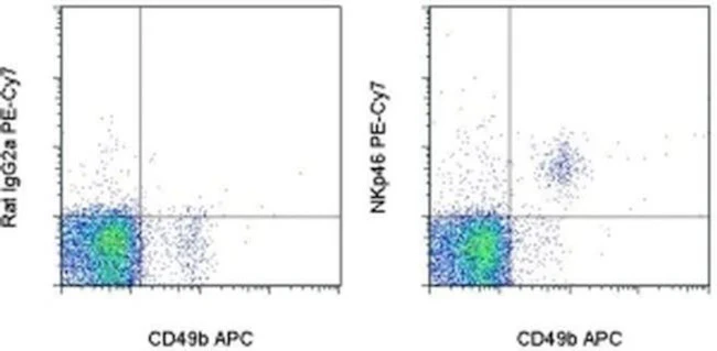
Description: The monoclonal antibody 29A1.4 recognizes mouse NKp46, also known as CD335. CD335, a member of the natural cytotoxicity receptor (NCR) family, is a glycoprotein with 2 Ig-like domains and a short cytoplasmic tail. Expression of CD335 is uniquely found on NK cells (including immature NK cells, defined as DX5- CD3-, and thereby allowing discrimination between NKT cells and NK cells (NKp46+, CD3-). Furthermore, unlike many of the NK markers which also stain NKT cells, staining with 29A1.4 is not strain specific. Staining has been shown on C57Bl/6, SJL, CBA/CA and BALB/C strains. NKp46 has been shown to play a role in NK cell-mediated lysis of several tumor cells and pathogen-infected cell lines.
The 29A1.4 monoclonal antibody has been shown to activate NK cells in vitro. The 29A1.4 monoclonal antibody does not deplete NK cells in vivo.
Applications Reported: This 29A1.4 antibody has been reported for use in flow cytometric analysis.
Applications Tested: This 29A1.4 antibody has been tested by flow cytometric analysis of mouse splenocytes. This can be used at less than or equal to 1 µg per test. A test is defined as the amount (µg) of antibody that will stain a cell sample in a final volume of 100 µL. Cell number should be determined empirically but can range from 10^5 to 10^8 cells/test. It is recommended that the antibody be carefully titrated for optimal performance in the assay of interest.
Light sensitivity: This tandem dye is sensitive photo-induced oxidation. Please protect this vial and stained samples from light.
Fixation: Samples can be stored in IC Fixation Buffer (Product # 00-8222) (100 µL cell sample + 100 µL IC Fixation Buffer) or 1-step Fix/Lyse Solution (Product # 00-5333) for up to 3 days in the dark at 4°C with minimal impact on brightness and FRET efficiency/compensation. Some generalizations regarding fluorophore performance after fixation can be made, but clone specific performance should be determined empirically.
Excitation: 488-561 nm; Emission: 775 nm; Laser: Blue Laser, Green Laser, Yellow-Green Laser.
Filtration: 0.2 µm post-manufacturing filtered.
For Research Use Only. Not for use in diagnostic procedures. Not for resale without express authorization.
CD335 (NKp46) Monoclonal Antibody (29A1.4), PE-Cyanine7, eBioscience
PRODUCT DETAILS
Host: Rat
Isotype: IgG2a, kappa
Clonality: Monoclonal
Clone: 29A1.4
Format: PE-Cyanine7
Reactivity: Ms
Application: Flow Cytometry
Tested Dilution: 1 µg/test
Concentration: 0.2 mg/mL
Storage: 4°C, store in dark, DO NOT FREEZE!
Formulation: PBS with 0.09% sodium azide; pH 7.2
Purification: Affinity chromatography
Data Sheet: TDS
Specific Information
Description: The monoclonal antibody 29A1.4 recognizes mouse NKp46, also known as CD335. CD335, a member of the natural cytotoxicity receptor (NCR) family, is a glycoprotein with 2 Ig-like domains and a short cytoplasmic tail. Expression of CD335 is uniquely found on NK cells (including immature NK cells, defined as DX5- CD3-, and thereby allowing discrimination between NKT cells and NK cells (NKp46+, CD3-). Furthermore, unlike many of the NK markers which also stain NKT cells, staining with 29A1.4 is not strain specific. Staining has been shown on C57Bl/6, SJL, CBA/CA and BALB/C strains. NKp46 has been shown to play a role in NK cell-mediated lysis of several tumor cells and pathogen-infected cell lines.
The 29A1.4 monoclonal antibody has been shown to activate NK cells in vitro. The 29A1.4 monoclonal antibody does not deplete NK cells in vivo.
Applications Reported: This 29A1.4 antibody has been reported for use in flow cytometric analysis.
Applications Tested: This 29A1.4 antibody has been tested by flow cytometric analysis of mouse splenocytes. This can be used at less than or equal to 1 µg per test. A test is defined as the amount (µg) of antibody that will stain a cell sample in a final volume of 100 µL. Cell number should be determined empirically but can range from 10^5 to 10^8 cells/test. It is recommended that the antibody be carefully titrated for optimal performance in the assay of interest.
Light sensitivity: This tandem dye is sensitive photo-induced oxidation. Please protect this vial and stained samples from light.
Fixation: Samples can be stored in IC Fixation Buffer (Product # 00-8222) (100 µL cell sample + 100 µL IC Fixation Buffer) or 1-step Fix/Lyse Solution (Product # 00-5333) for up to 3 days in the dark at 4°C with minimal impact on brightness and FRET efficiency/compensation. Some generalizations regarding fluorophore performance after fixation can be made, but clone specific performance should be determined empirically.
Excitation: 488-561 nm; Emission: 775 nm; Laser: Blue Laser, Green Laser, Yellow-Green Laser.
Filtration: 0.2 µm post-manufacturing filtered.
For Research Use Only. Not for use in diagnostic procedures. Not for resale without express authorization.
Product Information
Product Information
Shipping & Returns
Shipping & Returns
Description
PRODUCT DETAILS
Host: Rat
Isotype: IgG2a, kappa
Clonality: Monoclonal
Clone: 29A1.4
Format: PE-Cyanine7
Reactivity: Ms
Application: Flow Cytometry
Tested Dilution: 1 µg/test
Concentration: 0.2 mg/mL
Storage: 4°C, store in dark, DO NOT FREEZE!
Formulation: PBS with 0.09% sodium azide; pH 7.2
Purification: Affinity chromatography
Data Sheet: TDS
Specific Information
Description: The monoclonal antibody 29A1.4 recognizes mouse NKp46, also known as CD335. CD335, a member of the natural cytotoxicity receptor (NCR) family, is a glycoprotein with 2 Ig-like domains and a short cytoplasmic tail. Expression of CD335 is uniquely found on NK cells (including immature NK cells, defined as DX5- CD3-, and thereby allowing discrimination between NKT cells and NK cells (NKp46+, CD3-). Furthermore, unlike many of the NK markers which also stain NKT cells, staining with 29A1.4 is not strain specific. Staining has been shown on C57Bl/6, SJL, CBA/CA and BALB/C strains. NKp46 has been shown to play a role in NK cell-mediated lysis of several tumor cells and pathogen-infected cell lines.
The 29A1.4 monoclonal antibody has been shown to activate NK cells in vitro. The 29A1.4 monoclonal antibody does not deplete NK cells in vivo.
Applications Reported: This 29A1.4 antibody has been reported for use in flow cytometric analysis.
Applications Tested: This 29A1.4 antibody has been tested by flow cytometric analysis of mouse splenocytes. This can be used at less than or equal to 1 µg per test. A test is defined as the amount (µg) of antibody that will stain a cell sample in a final volume of 100 µL. Cell number should be determined empirically but can range from 10^5 to 10^8 cells/test. It is recommended that the antibody be carefully titrated for optimal performance in the assay of interest.
Light sensitivity: This tandem dye is sensitive photo-induced oxidation. Please protect this vial and stained samples from light.
Fixation: Samples can be stored in IC Fixation Buffer (Product # 00-8222) (100 µL cell sample + 100 µL IC Fixation Buffer) or 1-step Fix/Lyse Solution (Product # 00-5333) for up to 3 days in the dark at 4°C with minimal impact on brightness and FRET efficiency/compensation. Some generalizations regarding fluorophore performance after fixation can be made, but clone specific performance should be determined empirically.
Excitation: 488-561 nm; Emission: 775 nm; Laser: Blue Laser, Green Laser, Yellow-Green Laser.
Filtration: 0.2 µm post-manufacturing filtered.
For Research Use Only. Not for use in diagnostic procedures. Not for resale without express authorization.


