CD324 (E-Cadherin) Monoclonal Antibody (DECMA-1), Biotin, eBioscience
PRODUCT DETAILS
Host: Rat
Isotype: IgG1, kappa
Clonality: Monoclonal
Clone: DECMA-1
Format: Biotin
Reactivity: Ca, Hu, Ms
Application: Flow Cytometry
Tested Dilution: 0.25 µg/test
Concentration: 0.5 mg/mL
Storage: 4°C, store in dark, DO NOT FREEZE!
Formulation: PBS with 0.09% sodium azide; pH 7.2
Purification: Affinity chromatography
Data Sheet: TDS
Specific Information
Description: The monoclonal antibody DECMA-1 recognizes mouse, human and canine CD324 also known as E-cadherin (Epithelial cadherin) or uvomorulin. Like the other cadherin family members P and N cadherin, E-cadherin is a transmembrane glycoprotein involved in intercellular adhesion. These proteins share a common basic structure. The extracellular portions of the proteins are largely composed of repeating domains, each with two consensus Ca2+-binding motifs. The cytoplasmic domain interacts with a-, b-, and g-catenins and actinins. These catenins connect E-cadherin with the cytoskeleton.
Expression is found in most epidermal cells including melanocytes and kerotinocytes. E-cadherin is localized at the intercellular boundaries of epithelial cells in several tissues, and is thought to play a role in maintenance of tissue integrity. Loss of E-cadherin function has been implicated in the progression of a variety of cancers.
E-Cadherin protein is sensitive to trypsin treatment, so exposure to trypsin should be minimized or avoided.
The monoclonal antibody DECMA-1 has been shown to have functional activity by disrupting adhesion in human, mouse and dog cells.
Applications Reported: This DECMA-1 antibody has been reported for use in flow cytometric analysis, western blotting, and immunohistochemical staining of frozen tissue sections.
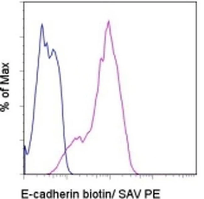
Applications Tested: This DECMA-1 antibody has been tested by flow cytometric analysis of the MDCK cell line. Optimal staining is achieved by intracellular staining as protein turnover can result in variable surface staining. This can be used at less than or equal to 0.25 µg per test. A test is defined as the amount (µg) of antibody that will stain a cell sample in a final volume of 100 µL. Cell number should be determined empirically but can range from 10^5 to 10^8 cells/test. It is recommended that the antibody be carefully titrated for optimal performance in the assay of interest.
Filtration: 0.2 µm post-manufacturing filtered.
For Research Use Only. Not for use in diagnostic procedures. Not for resale without express authorization.
CD324 (E-Cadherin) Monoclonal Antibody (DECMA-1), Biotin, eBioscience
PRODUCT DETAILS
Host: Rat
Isotype: IgG1, kappa
Clonality: Monoclonal
Clone: DECMA-1
Format: Biotin
Reactivity: Ca, Hu, Ms
Application: Flow Cytometry
Tested Dilution: 0.25 µg/test
Concentration: 0.5 mg/mL
Storage: 4°C, store in dark, DO NOT FREEZE!
Formulation: PBS with 0.09% sodium azide; pH 7.2
Purification: Affinity chromatography
Data Sheet: TDS
Specific Information
Description: The monoclonal antibody DECMA-1 recognizes mouse, human and canine CD324 also known as E-cadherin (Epithelial cadherin) or uvomorulin. Like the other cadherin family members P and N cadherin, E-cadherin is a transmembrane glycoprotein involved in intercellular adhesion. These proteins share a common basic structure. The extracellular portions of the proteins are largely composed of repeating domains, each with two consensus Ca2+-binding motifs. The cytoplasmic domain interacts with a-, b-, and g-catenins and actinins. These catenins connect E-cadherin with the cytoskeleton.
Expression is found in most epidermal cells including melanocytes and kerotinocytes. E-cadherin is localized at the intercellular boundaries of epithelial cells in several tissues, and is thought to play a role in maintenance of tissue integrity. Loss of E-cadherin function has been implicated in the progression of a variety of cancers.
E-Cadherin protein is sensitive to trypsin treatment, so exposure to trypsin should be minimized or avoided.
The monoclonal antibody DECMA-1 has been shown to have functional activity by disrupting adhesion in human, mouse and dog cells.
Applications Reported: This DECMA-1 antibody has been reported for use in flow cytometric analysis, western blotting, and immunohistochemical staining of frozen tissue sections.
Applications Tested: This DECMA-1 antibody has been tested by flow cytometric analysis of the MDCK cell line. Optimal staining is achieved by intracellular staining as protein turnover can result in variable surface staining. This can be used at less than or equal to 0.25 µg per test. A test is defined as the amount (µg) of antibody that will stain a cell sample in a final volume of 100 µL. Cell number should be determined empirically but can range from 10^5 to 10^8 cells/test. It is recommended that the antibody be carefully titrated for optimal performance in the assay of interest.
Filtration: 0.2 µm post-manufacturing filtered.
For Research Use Only. Not for use in diagnostic procedures. Not for resale without express authorization.
Product Information
Product Information
Shipping & Returns
Shipping & Returns
Description
PRODUCT DETAILS
Host: Rat
Isotype: IgG1, kappa
Clonality: Monoclonal
Clone: DECMA-1
Format: Biotin
Reactivity: Ca, Hu, Ms
Application: Flow Cytometry
Tested Dilution: 0.25 µg/test
Concentration: 0.5 mg/mL
Storage: 4°C, store in dark, DO NOT FREEZE!
Formulation: PBS with 0.09% sodium azide; pH 7.2
Purification: Affinity chromatography
Data Sheet: TDS
Specific Information
Description: The monoclonal antibody DECMA-1 recognizes mouse, human and canine CD324 also known as E-cadherin (Epithelial cadherin) or uvomorulin. Like the other cadherin family members P and N cadherin, E-cadherin is a transmembrane glycoprotein involved in intercellular adhesion. These proteins share a common basic structure. The extracellular portions of the proteins are largely composed of repeating domains, each with two consensus Ca2+-binding motifs. The cytoplasmic domain interacts with a-, b-, and g-catenins and actinins. These catenins connect E-cadherin with the cytoskeleton.
Expression is found in most epidermal cells including melanocytes and kerotinocytes. E-cadherin is localized at the intercellular boundaries of epithelial cells in several tissues, and is thought to play a role in maintenance of tissue integrity. Loss of E-cadherin function has been implicated in the progression of a variety of cancers.
E-Cadherin protein is sensitive to trypsin treatment, so exposure to trypsin should be minimized or avoided.
The monoclonal antibody DECMA-1 has been shown to have functional activity by disrupting adhesion in human, mouse and dog cells.
Applications Reported: This DECMA-1 antibody has been reported for use in flow cytometric analysis, western blotting, and immunohistochemical staining of frozen tissue sections.
Applications Tested: This DECMA-1 antibody has been tested by flow cytometric analysis of the MDCK cell line. Optimal staining is achieved by intracellular staining as protein turnover can result in variable surface staining. This can be used at less than or equal to 0.25 µg per test. A test is defined as the amount (µg) of antibody that will stain a cell sample in a final volume of 100 µL. Cell number should be determined empirically but can range from 10^5 to 10^8 cells/test. It is recommended that the antibody be carefully titrated for optimal performance in the assay of interest.
Filtration: 0.2 µm post-manufacturing filtered.
For Research Use Only. Not for use in diagnostic procedures. Not for resale without express authorization.


