CD294 (CRTH2) Monoclonal Antibody (No3m1scz), Alexa Fluor 647, eBioscience
PRODUCT DETAILS
Host: Rat
Isotype: IgG2a, kappa
Clonality: Monoclonal
Clone: No3m1scz
Format: Alexa Fluor 647
Reactivity: Ms
Application: Flow Cytometry
Tested Dilution: 0.25 µg
Concentration: 0.2 mg/mL
Storage: 4°C, store in dark, DO NOT FREEZE!
Formulation: PBS with BSA and 0.09% sodium azide; pH 7.2
Purification: Affinity chromatography
Data Sheet: TDS
Specific Information
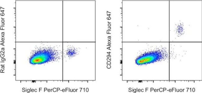
Description: This clone recognizes mouse CD294, also known as Prostaglandin D2 receptor 2 (PD2R2), G-protein coupled receptor 44 (GPR44) or Chemoattractant Receptor-Homologous Molecule Expressed On TH2 Cells (CRTH2). Although the clone works on unfixed mouse CD294-transfected cells, we recommend fixing and permeabilizing primary cells.
Applications Reported: This No3m1scz antibody has been reported for use in flow cytometric analysis.
Applications Tested: This No3m1scz antibody has been tested by flow cytometric analysis of mouse lysed whole blood cells using Protocol A: Two-step protocol for intracellular (cytoplasmic) proteins. This allows for the greatest flexibility for detection of surface and intracellular (cytoplasmic) proteins. Use of Protocol B: One-step protocol for intracellular (nuclear) proteins is recommended for staining of transcription factors in conjunction with surface and phosphorylated intracellular (cytoplasmic) proteins. Protocol C: Two-step protocol: Fixation/Methanol allows for the greatest discrimination of phospho-specific signaling between unstimulated and stimulated samples, but with limitations on the ability to stain specific surface proteins (refer to ″Clone Performance Following Fixation/Permeabilization″). All Protocols can be found in the ″Staining Intracellular Antigens for Flow Cytometry Protocol″ located in the BestProtocols® Section under the Resources tab online. This may be used at less than or equal to 0.25 µg per test. A test is defined as the amount (µg) of antibody that will stain a cell sample in a final volume of 100 µL. Cell number should be determined empirically but can range from 10^5 to 10^8 cells/test. It is recommended that the antibody be carefully titrated for optimal performance in the assay of interest.
Excitation: 633-647 nm; Emission: 668 nm; Laser: Red Laser
For Research Use Only. Not for use in diagnostic procedures. Not for resale without express authorization.
Original: $506.00
-70%$506.00
$151.80CD294 (CRTH2) Monoclonal Antibody (No3m1scz), Alexa Fluor 647, eBioscience
PRODUCT DETAILS
Host: Rat
Isotype: IgG2a, kappa
Clonality: Monoclonal
Clone: No3m1scz
Format: Alexa Fluor 647
Reactivity: Ms
Application: Flow Cytometry
Tested Dilution: 0.25 µg
Concentration: 0.2 mg/mL
Storage: 4°C, store in dark, DO NOT FREEZE!
Formulation: PBS with BSA and 0.09% sodium azide; pH 7.2
Purification: Affinity chromatography
Data Sheet: TDS
Specific Information
Description: This clone recognizes mouse CD294, also known as Prostaglandin D2 receptor 2 (PD2R2), G-protein coupled receptor 44 (GPR44) or Chemoattractant Receptor-Homologous Molecule Expressed On TH2 Cells (CRTH2). Although the clone works on unfixed mouse CD294-transfected cells, we recommend fixing and permeabilizing primary cells.
Applications Reported: This No3m1scz antibody has been reported for use in flow cytometric analysis.
Applications Tested: This No3m1scz antibody has been tested by flow cytometric analysis of mouse lysed whole blood cells using Protocol A: Two-step protocol for intracellular (cytoplasmic) proteins. This allows for the greatest flexibility for detection of surface and intracellular (cytoplasmic) proteins. Use of Protocol B: One-step protocol for intracellular (nuclear) proteins is recommended for staining of transcription factors in conjunction with surface and phosphorylated intracellular (cytoplasmic) proteins. Protocol C: Two-step protocol: Fixation/Methanol allows for the greatest discrimination of phospho-specific signaling between unstimulated and stimulated samples, but with limitations on the ability to stain specific surface proteins (refer to ″Clone Performance Following Fixation/Permeabilization″). All Protocols can be found in the ″Staining Intracellular Antigens for Flow Cytometry Protocol″ located in the BestProtocols® Section under the Resources tab online. This may be used at less than or equal to 0.25 µg per test. A test is defined as the amount (µg) of antibody that will stain a cell sample in a final volume of 100 µL. Cell number should be determined empirically but can range from 10^5 to 10^8 cells/test. It is recommended that the antibody be carefully titrated for optimal performance in the assay of interest.
Excitation: 633-647 nm; Emission: 668 nm; Laser: Red Laser
For Research Use Only. Not for use in diagnostic procedures. Not for resale without express authorization.
Product Information
Product Information
Shipping & Returns
Shipping & Returns
Description
PRODUCT DETAILS
Host: Rat
Isotype: IgG2a, kappa
Clonality: Monoclonal
Clone: No3m1scz
Format: Alexa Fluor 647
Reactivity: Ms
Application: Flow Cytometry
Tested Dilution: 0.25 µg
Concentration: 0.2 mg/mL
Storage: 4°C, store in dark, DO NOT FREEZE!
Formulation: PBS with BSA and 0.09% sodium azide; pH 7.2
Purification: Affinity chromatography
Data Sheet: TDS
Specific Information
Description: This clone recognizes mouse CD294, also known as Prostaglandin D2 receptor 2 (PD2R2), G-protein coupled receptor 44 (GPR44) or Chemoattractant Receptor-Homologous Molecule Expressed On TH2 Cells (CRTH2). Although the clone works on unfixed mouse CD294-transfected cells, we recommend fixing and permeabilizing primary cells.
Applications Reported: This No3m1scz antibody has been reported for use in flow cytometric analysis.
Applications Tested: This No3m1scz antibody has been tested by flow cytometric analysis of mouse lysed whole blood cells using Protocol A: Two-step protocol for intracellular (cytoplasmic) proteins. This allows for the greatest flexibility for detection of surface and intracellular (cytoplasmic) proteins. Use of Protocol B: One-step protocol for intracellular (nuclear) proteins is recommended for staining of transcription factors in conjunction with surface and phosphorylated intracellular (cytoplasmic) proteins. Protocol C: Two-step protocol: Fixation/Methanol allows for the greatest discrimination of phospho-specific signaling between unstimulated and stimulated samples, but with limitations on the ability to stain specific surface proteins (refer to ″Clone Performance Following Fixation/Permeabilization″). All Protocols can be found in the ″Staining Intracellular Antigens for Flow Cytometry Protocol″ located in the BestProtocols® Section under the Resources tab online. This may be used at less than or equal to 0.25 µg per test. A test is defined as the amount (µg) of antibody that will stain a cell sample in a final volume of 100 µL. Cell number should be determined empirically but can range from 10^5 to 10^8 cells/test. It is recommended that the antibody be carefully titrated for optimal performance in the assay of interest.
Excitation: 633-647 nm; Emission: 668 nm; Laser: Red Laser
For Research Use Only. Not for use in diagnostic procedures. Not for resale without express authorization.


