CD279 (PD-1) Monoclonal Antibody (J43), PerCP-eFluor 710, eBioscience
PRODUCT DETAILS
Host: Armenian Hamster
Isotype: IgG
Clonality: Monoclonal
Clone: J43
Format: PerCP-eFluor 710
Reactivity: Ms
Application: Flow Cytometry
Tested Dilution: 0.125 µg/test
Concentration: 0.2 mg/mL
Storage: 4°C, store in dark, DO NOT FREEZE!
Formulation: PBS with 0.09% sodium azide; pH 7.2
Purification: Affinity chromatography
Data Sheet: TDS
Specific Information
Description: The J43 monoclonal antibody reacts with mouse PD-1 (programmed death-1), a 55 kDa member of the Ig superfamily. PD-1 contains the immunoreceptor tyrosine-based inhibitory motif (ITIM) and plays a key role in peripheral tolerance and autoimmune disease in mice. PD-1 is expressed mainly on activated T and B lymphocytes. Two novel B7 Family members have been identified as PD-1 ligands, PD-L1 (B7-H1) and PD-L2 (B7-DC). Evidence reported to date suggests overlapping functions for these ligands and their constitutive expression on some normal tissues and upregulation on activated antigen-presenting cells. It is reported that J43 inhibits the binding of mouse PD-L1-Ig and mouse PD-L2-Ig to PD-1/BHK transfected cells. When administrated in vivo, both intact and Fab of J43 are reported to enhance contact hypersensitivity and exacerbate acute GVHD similar to transfer of PD-1-deficient cells. Injection of J43 also exacerbates EAE and NOD diabetes as do specific antibodies to mouse PD-L1 and PD-L2.
Applications Reported: This J43 antibody has been reported for use in flow cytometric analysis.
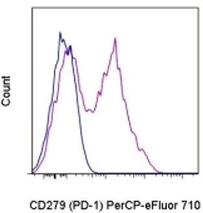
Applications Tested: This J43 antibody has been tested by flow cytometric analysis of stimulated mouse splenocytes. This can be used at less than or equal to 0.125 µg per test. A test is defined as the amount (µg) of antibody that will stain a cell sample in a final volume of 100 µL. Cell number should be determined empirically but can range from 10^5 to 10^8 cells/test. It is recommended that the antibody be carefully titrated for optimal performance in the assay of interest.
PerCP-eFluor® 710 can be used in place of PE-Cy5, PE-Cy5.5 or PerCP-Cy5.5. PerCP-eFluor® 710 emits at 710 nm and is excited with the blue laser (488 nm). Please make sure that your instrument is capable of detecting this fluorochrome. For a filter configuration, we recommend using the 685 LP dichroic mirror and 710/40 band pass filter, however the 695/40 band pass filter is an acceptable alternative.
Our testing indicates that PerCP-eFluor® 710 conjugated antibodies are stable when stained samples are exposed to freshly prepared 2% formaldehyde overnight at 4°C, but please evaluate for alternative fixation protocols.
Excitation: 488 nm; Emission: 710 nm; Laser: Blue Laser.
Filtration: 0.2 µm post-manufacturing filtered.
For Research Use Only. Not for use in diagnostic procedures. Not for resale without express authorization.
Original: $447.00
-70%$447.00
$134.10CD279 (PD-1) Monoclonal Antibody (J43), PerCP-eFluor 710, eBioscience
PRODUCT DETAILS
Host: Armenian Hamster
Isotype: IgG
Clonality: Monoclonal
Clone: J43
Format: PerCP-eFluor 710
Reactivity: Ms
Application: Flow Cytometry
Tested Dilution: 0.125 µg/test
Concentration: 0.2 mg/mL
Storage: 4°C, store in dark, DO NOT FREEZE!
Formulation: PBS with 0.09% sodium azide; pH 7.2
Purification: Affinity chromatography
Data Sheet: TDS
Specific Information
Description: The J43 monoclonal antibody reacts with mouse PD-1 (programmed death-1), a 55 kDa member of the Ig superfamily. PD-1 contains the immunoreceptor tyrosine-based inhibitory motif (ITIM) and plays a key role in peripheral tolerance and autoimmune disease in mice. PD-1 is expressed mainly on activated T and B lymphocytes. Two novel B7 Family members have been identified as PD-1 ligands, PD-L1 (B7-H1) and PD-L2 (B7-DC). Evidence reported to date suggests overlapping functions for these ligands and their constitutive expression on some normal tissues and upregulation on activated antigen-presenting cells. It is reported that J43 inhibits the binding of mouse PD-L1-Ig and mouse PD-L2-Ig to PD-1/BHK transfected cells. When administrated in vivo, both intact and Fab of J43 are reported to enhance contact hypersensitivity and exacerbate acute GVHD similar to transfer of PD-1-deficient cells. Injection of J43 also exacerbates EAE and NOD diabetes as do specific antibodies to mouse PD-L1 and PD-L2.
Applications Reported: This J43 antibody has been reported for use in flow cytometric analysis.
Applications Tested: This J43 antibody has been tested by flow cytometric analysis of stimulated mouse splenocytes. This can be used at less than or equal to 0.125 µg per test. A test is defined as the amount (µg) of antibody that will stain a cell sample in a final volume of 100 µL. Cell number should be determined empirically but can range from 10^5 to 10^8 cells/test. It is recommended that the antibody be carefully titrated for optimal performance in the assay of interest.
PerCP-eFluor® 710 can be used in place of PE-Cy5, PE-Cy5.5 or PerCP-Cy5.5. PerCP-eFluor® 710 emits at 710 nm and is excited with the blue laser (488 nm). Please make sure that your instrument is capable of detecting this fluorochrome. For a filter configuration, we recommend using the 685 LP dichroic mirror and 710/40 band pass filter, however the 695/40 band pass filter is an acceptable alternative.
Our testing indicates that PerCP-eFluor® 710 conjugated antibodies are stable when stained samples are exposed to freshly prepared 2% formaldehyde overnight at 4°C, but please evaluate for alternative fixation protocols.
Excitation: 488 nm; Emission: 710 nm; Laser: Blue Laser.
Filtration: 0.2 µm post-manufacturing filtered.
For Research Use Only. Not for use in diagnostic procedures. Not for resale without express authorization.
Product Information
Product Information
Shipping & Returns
Shipping & Returns
Description
PRODUCT DETAILS
Host: Armenian Hamster
Isotype: IgG
Clonality: Monoclonal
Clone: J43
Format: PerCP-eFluor 710
Reactivity: Ms
Application: Flow Cytometry
Tested Dilution: 0.125 µg/test
Concentration: 0.2 mg/mL
Storage: 4°C, store in dark, DO NOT FREEZE!
Formulation: PBS with 0.09% sodium azide; pH 7.2
Purification: Affinity chromatography
Data Sheet: TDS
Specific Information
Description: The J43 monoclonal antibody reacts with mouse PD-1 (programmed death-1), a 55 kDa member of the Ig superfamily. PD-1 contains the immunoreceptor tyrosine-based inhibitory motif (ITIM) and plays a key role in peripheral tolerance and autoimmune disease in mice. PD-1 is expressed mainly on activated T and B lymphocytes. Two novel B7 Family members have been identified as PD-1 ligands, PD-L1 (B7-H1) and PD-L2 (B7-DC). Evidence reported to date suggests overlapping functions for these ligands and their constitutive expression on some normal tissues and upregulation on activated antigen-presenting cells. It is reported that J43 inhibits the binding of mouse PD-L1-Ig and mouse PD-L2-Ig to PD-1/BHK transfected cells. When administrated in vivo, both intact and Fab of J43 are reported to enhance contact hypersensitivity and exacerbate acute GVHD similar to transfer of PD-1-deficient cells. Injection of J43 also exacerbates EAE and NOD diabetes as do specific antibodies to mouse PD-L1 and PD-L2.
Applications Reported: This J43 antibody has been reported for use in flow cytometric analysis.
Applications Tested: This J43 antibody has been tested by flow cytometric analysis of stimulated mouse splenocytes. This can be used at less than or equal to 0.125 µg per test. A test is defined as the amount (µg) of antibody that will stain a cell sample in a final volume of 100 µL. Cell number should be determined empirically but can range from 10^5 to 10^8 cells/test. It is recommended that the antibody be carefully titrated for optimal performance in the assay of interest.
PerCP-eFluor® 710 can be used in place of PE-Cy5, PE-Cy5.5 or PerCP-Cy5.5. PerCP-eFluor® 710 emits at 710 nm and is excited with the blue laser (488 nm). Please make sure that your instrument is capable of detecting this fluorochrome. For a filter configuration, we recommend using the 685 LP dichroic mirror and 710/40 band pass filter, however the 695/40 band pass filter is an acceptable alternative.
Our testing indicates that PerCP-eFluor® 710 conjugated antibodies are stable when stained samples are exposed to freshly prepared 2% formaldehyde overnight at 4°C, but please evaluate for alternative fixation protocols.
Excitation: 488 nm; Emission: 710 nm; Laser: Blue Laser.
Filtration: 0.2 µm post-manufacturing filtered.
For Research Use Only. Not for use in diagnostic procedures. Not for resale without express authorization.


