CD279 (PD-1) Monoclonal Antibody (J43), Functional Grade, eBioscience
PRODUCT DETAILS
Host: Armenian Hamster
Isotype: IgG
Clonality: Monoclonal
Clone: J43
Format: Functional Grade
Reactivity: Ms
Application: Flow Cytometry
Tested Dilution: 0.5 µg/test
Concentration: 1 mg/mL
Storage: 4°C
Formulation: PBS with no preservative; pH 7.2
Purification: Affinity chromatography
Data Sheet: TDS
Specific Information
Description: The J43 monoclonal antibody reacts with mouse PD-1 (programmed death-1), a 55 kDa member of the Ig superfamily. PD-1 contains the immunoreceptor tyrosine-based inhibitory motif (ITIM) and plays a key role in peripheral tolerance and autoimmune disease in mice. PD-1 is expressed mainly on activated T and B lymphocytes. Two novel B7 Family members have been identified as PD-1 ligands, PD-L1 (B7-H1) and PD-L2 (B7-DC). Evidence reported to date suggests overlapping functions for these ligands and their constitutive expression on some normal tissues and upregulation on activated antigen-presenting cells. It is reported that J43 inhibits the binding of mouse PD-L1-Ig and mouse PD-L2-Ig to PD-1/BHK transfected cells. When administrated in vivo, both intact and Fab of J43 are reported to enhance contact hypersensitivity and exacerbate acute GVHD similar to transfer of PD-1-deficient cells. Injection of J43 also exacerbates EAE and NOD diabetes as do specific antibodies to mouse PD-L1 and PD-L2.
Applications Reported: The J43 antibody has been reported for use in flow cytometric analysis. It has also been reported for use in in vitro functional assays.
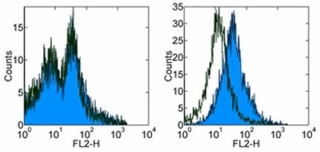
Applications Tested: The J43 antibody has been tested by flow cytometric analysis of mouse Con-A activated spleen cells and mouse PD-1 transfected cells. This can be used at less than or equal to 0.5 µg per test. A test is defined as the amount (µg) of antibody that will stain a cell sample in a final volume of 100 µL. Cell number should be determined empirically but can range from 10^5 to 10^8 cells/test. It is recommended that the antibody be carefully titrated for optimal performance in the assay of interest.
Storage and handling: Use in a sterile environment.
Filtration: 0.2 µm post-manufacturing filtered.
Purity: Greater than 90%, as determined by SDS-PAGE.
Endotoxin Level: Less than 0.001 ng/µg antibody, as determined by LAL assay.
Aggregation: Less than 10%, as determined by HPLC.
For Research Use Only. Not for use in diagnostic procedures. Not for resale without express authorization.
CD279 (PD-1) Monoclonal Antibody (J43), Functional Grade, eBioscience
PRODUCT DETAILS
Host: Armenian Hamster
Isotype: IgG
Clonality: Monoclonal
Clone: J43
Format: Functional Grade
Reactivity: Ms
Application: Flow Cytometry
Tested Dilution: 0.5 µg/test
Concentration: 1 mg/mL
Storage: 4°C
Formulation: PBS with no preservative; pH 7.2
Purification: Affinity chromatography
Data Sheet: TDS
Specific Information
Description: The J43 monoclonal antibody reacts with mouse PD-1 (programmed death-1), a 55 kDa member of the Ig superfamily. PD-1 contains the immunoreceptor tyrosine-based inhibitory motif (ITIM) and plays a key role in peripheral tolerance and autoimmune disease in mice. PD-1 is expressed mainly on activated T and B lymphocytes. Two novel B7 Family members have been identified as PD-1 ligands, PD-L1 (B7-H1) and PD-L2 (B7-DC). Evidence reported to date suggests overlapping functions for these ligands and their constitutive expression on some normal tissues and upregulation on activated antigen-presenting cells. It is reported that J43 inhibits the binding of mouse PD-L1-Ig and mouse PD-L2-Ig to PD-1/BHK transfected cells. When administrated in vivo, both intact and Fab of J43 are reported to enhance contact hypersensitivity and exacerbate acute GVHD similar to transfer of PD-1-deficient cells. Injection of J43 also exacerbates EAE and NOD diabetes as do specific antibodies to mouse PD-L1 and PD-L2.
Applications Reported: The J43 antibody has been reported for use in flow cytometric analysis. It has also been reported for use in in vitro functional assays.
Applications Tested: The J43 antibody has been tested by flow cytometric analysis of mouse Con-A activated spleen cells and mouse PD-1 transfected cells. This can be used at less than or equal to 0.5 µg per test. A test is defined as the amount (µg) of antibody that will stain a cell sample in a final volume of 100 µL. Cell number should be determined empirically but can range from 10^5 to 10^8 cells/test. It is recommended that the antibody be carefully titrated for optimal performance in the assay of interest.
Storage and handling: Use in a sterile environment.
Filtration: 0.2 µm post-manufacturing filtered.
Purity: Greater than 90%, as determined by SDS-PAGE.
Endotoxin Level: Less than 0.001 ng/µg antibody, as determined by LAL assay.
Aggregation: Less than 10%, as determined by HPLC.
For Research Use Only. Not for use in diagnostic procedures. Not for resale without express authorization.
Product Information
Product Information
Shipping & Returns
Shipping & Returns
Description
PRODUCT DETAILS
Host: Armenian Hamster
Isotype: IgG
Clonality: Monoclonal
Clone: J43
Format: Functional Grade
Reactivity: Ms
Application: Flow Cytometry
Tested Dilution: 0.5 µg/test
Concentration: 1 mg/mL
Storage: 4°C
Formulation: PBS with no preservative; pH 7.2
Purification: Affinity chromatography
Data Sheet: TDS
Specific Information
Description: The J43 monoclonal antibody reacts with mouse PD-1 (programmed death-1), a 55 kDa member of the Ig superfamily. PD-1 contains the immunoreceptor tyrosine-based inhibitory motif (ITIM) and plays a key role in peripheral tolerance and autoimmune disease in mice. PD-1 is expressed mainly on activated T and B lymphocytes. Two novel B7 Family members have been identified as PD-1 ligands, PD-L1 (B7-H1) and PD-L2 (B7-DC). Evidence reported to date suggests overlapping functions for these ligands and their constitutive expression on some normal tissues and upregulation on activated antigen-presenting cells. It is reported that J43 inhibits the binding of mouse PD-L1-Ig and mouse PD-L2-Ig to PD-1/BHK transfected cells. When administrated in vivo, both intact and Fab of J43 are reported to enhance contact hypersensitivity and exacerbate acute GVHD similar to transfer of PD-1-deficient cells. Injection of J43 also exacerbates EAE and NOD diabetes as do specific antibodies to mouse PD-L1 and PD-L2.
Applications Reported: The J43 antibody has been reported for use in flow cytometric analysis. It has also been reported for use in in vitro functional assays.
Applications Tested: The J43 antibody has been tested by flow cytometric analysis of mouse Con-A activated spleen cells and mouse PD-1 transfected cells. This can be used at less than or equal to 0.5 µg per test. A test is defined as the amount (µg) of antibody that will stain a cell sample in a final volume of 100 µL. Cell number should be determined empirically but can range from 10^5 to 10^8 cells/test. It is recommended that the antibody be carefully titrated for optimal performance in the assay of interest.
Storage and handling: Use in a sterile environment.
Filtration: 0.2 µm post-manufacturing filtered.
Purity: Greater than 90%, as determined by SDS-PAGE.
Endotoxin Level: Less than 0.001 ng/µg antibody, as determined by LAL assay.
Aggregation: Less than 10%, as determined by HPLC.
For Research Use Only. Not for use in diagnostic procedures. Not for resale without express authorization.


