CD279 (PD-1) Monoclonal Antibody (J43), eFluor 506, eBioscience
PRODUCT DETAILS
Host: Armenian Hamster
Isotype: IgG
Clonality: Monoclonal
Clone: J43
Format: eFluor 506
Reactivity: Ms
Application: Flow Cytometry
Tested Dilution: 1 µg/test
Concentration: 0.2 mg/mL
Storage: 4°C, store in dark, DO NOT FREEZE!
Formulation: PBS with 0.09% sodium azide
Purification: Affinity chromatography
Data Sheet: TDS
Specific Information
The J43 monoclonal antibody reacts with mouse PD-1 (programmed death-1), a 55 kDa member of the Ig superfamily. PD-1 contains the immunoreceptor tyrosine-based inhibitory motif (ITIM) and plays a key role in peripheral tolerance and autoimmune disease in mice. PD-1 is expressed mainly on activated T and B lymphocytes. Two novel B7 Family members have been identified as PD-1 ligands, PD-L1 (B7-H1) and PD-L2 (B7-DC). Evidence reported to date suggests overlapping functions for these ligands and their constitutive expression on some normal tissues and upregulation on activated antigen-presenting cells. It is reported that J43 inhibits the binding of mouse PD-L1-Ig and mouse PD-L2-Ig to PD-1/BHK transfected cells. When administrated in vivo, both intact and Fab of J43 are reported to enhance contact hypersensitivity and exacerbate acute GVHD similar to transfer of PD-1- deficient cells. Injection of J43 also exacerbates EAE and NOD diabetes as do specific antibodies to mouse PD-L1 and PD-L2.
The J43 antibody has been reported for use in flow cytometric analysis.
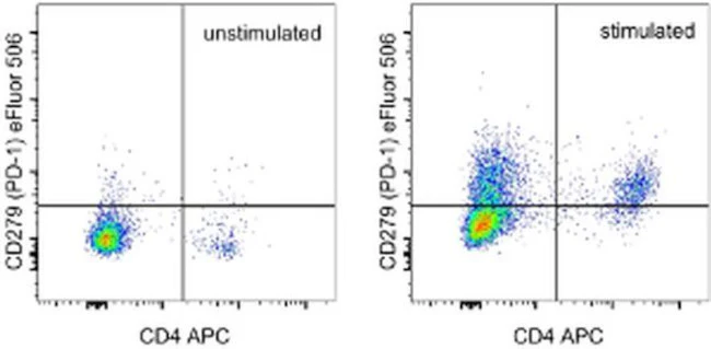
This J43 antibody has been tested by flow cytometric analysis of stimulated mouse splenocytes. This can be used at less than or equal to 1 µg per test. A test is defined as the amount (µg) of antibody that will stain a cell sample in a final volume of 100 µL. Cell number should be determined empirically but can range from 105 to 108 cells/test. It is recommended that the antibody be carefully titrated for optimal performance in the assay of interest.
eFluor™ 506 can be excited with the violet laser line (405 nm) and emits at 506 nm. We recommend using a 510/20 band pass filter, or equivalent. Please make sure that your instrument is capable of detecting this fluorochrome.
Filtration: 0.2 µm post-manufacturing filtered.
For Research Use Only. Not for use in diagnostic procedures. Not for resale without express authorization.
Original: $361.00
-70%$361.00
$108.30CD279 (PD-1) Monoclonal Antibody (J43), eFluor 506, eBioscience
PRODUCT DETAILS
Host: Armenian Hamster
Isotype: IgG
Clonality: Monoclonal
Clone: J43
Format: eFluor 506
Reactivity: Ms
Application: Flow Cytometry
Tested Dilution: 1 µg/test
Concentration: 0.2 mg/mL
Storage: 4°C, store in dark, DO NOT FREEZE!
Formulation: PBS with 0.09% sodium azide
Purification: Affinity chromatography
Data Sheet: TDS
Specific Information
The J43 monoclonal antibody reacts with mouse PD-1 (programmed death-1), a 55 kDa member of the Ig superfamily. PD-1 contains the immunoreceptor tyrosine-based inhibitory motif (ITIM) and plays a key role in peripheral tolerance and autoimmune disease in mice. PD-1 is expressed mainly on activated T and B lymphocytes. Two novel B7 Family members have been identified as PD-1 ligands, PD-L1 (B7-H1) and PD-L2 (B7-DC). Evidence reported to date suggests overlapping functions for these ligands and their constitutive expression on some normal tissues and upregulation on activated antigen-presenting cells. It is reported that J43 inhibits the binding of mouse PD-L1-Ig and mouse PD-L2-Ig to PD-1/BHK transfected cells. When administrated in vivo, both intact and Fab of J43 are reported to enhance contact hypersensitivity and exacerbate acute GVHD similar to transfer of PD-1- deficient cells. Injection of J43 also exacerbates EAE and NOD diabetes as do specific antibodies to mouse PD-L1 and PD-L2.
The J43 antibody has been reported for use in flow cytometric analysis.
This J43 antibody has been tested by flow cytometric analysis of stimulated mouse splenocytes. This can be used at less than or equal to 1 µg per test. A test is defined as the amount (µg) of antibody that will stain a cell sample in a final volume of 100 µL. Cell number should be determined empirically but can range from 105 to 108 cells/test. It is recommended that the antibody be carefully titrated for optimal performance in the assay of interest.
eFluor™ 506 can be excited with the violet laser line (405 nm) and emits at 506 nm. We recommend using a 510/20 band pass filter, or equivalent. Please make sure that your instrument is capable of detecting this fluorochrome.
Filtration: 0.2 µm post-manufacturing filtered.
For Research Use Only. Not for use in diagnostic procedures. Not for resale without express authorization.
Product Information
Product Information
Shipping & Returns
Shipping & Returns
Description
PRODUCT DETAILS
Host: Armenian Hamster
Isotype: IgG
Clonality: Monoclonal
Clone: J43
Format: eFluor 506
Reactivity: Ms
Application: Flow Cytometry
Tested Dilution: 1 µg/test
Concentration: 0.2 mg/mL
Storage: 4°C, store in dark, DO NOT FREEZE!
Formulation: PBS with 0.09% sodium azide
Purification: Affinity chromatography
Data Sheet: TDS
Specific Information
The J43 monoclonal antibody reacts with mouse PD-1 (programmed death-1), a 55 kDa member of the Ig superfamily. PD-1 contains the immunoreceptor tyrosine-based inhibitory motif (ITIM) and plays a key role in peripheral tolerance and autoimmune disease in mice. PD-1 is expressed mainly on activated T and B lymphocytes. Two novel B7 Family members have been identified as PD-1 ligands, PD-L1 (B7-H1) and PD-L2 (B7-DC). Evidence reported to date suggests overlapping functions for these ligands and their constitutive expression on some normal tissues and upregulation on activated antigen-presenting cells. It is reported that J43 inhibits the binding of mouse PD-L1-Ig and mouse PD-L2-Ig to PD-1/BHK transfected cells. When administrated in vivo, both intact and Fab of J43 are reported to enhance contact hypersensitivity and exacerbate acute GVHD similar to transfer of PD-1- deficient cells. Injection of J43 also exacerbates EAE and NOD diabetes as do specific antibodies to mouse PD-L1 and PD-L2.
The J43 antibody has been reported for use in flow cytometric analysis.
This J43 antibody has been tested by flow cytometric analysis of stimulated mouse splenocytes. This can be used at less than or equal to 1 µg per test. A test is defined as the amount (µg) of antibody that will stain a cell sample in a final volume of 100 µL. Cell number should be determined empirically but can range from 105 to 108 cells/test. It is recommended that the antibody be carefully titrated for optimal performance in the assay of interest.
eFluor™ 506 can be excited with the violet laser line (405 nm) and emits at 506 nm. We recommend using a 510/20 band pass filter, or equivalent. Please make sure that your instrument is capable of detecting this fluorochrome.
Filtration: 0.2 µm post-manufacturing filtered.
For Research Use Only. Not for use in diagnostic procedures. Not for resale without express authorization.


