CD279 (PD-1) Monoclonal Antibody (J43), Brilliant Violet 786, eBioscience
PRODUCT DETAILS
Host: Armenian Hamster
Isotype: IgG
Clonality: Monoclonal
Clone: J43
Format: Brilliant Violet 786
Reactivity: Ms
Application: Flow Cytometry
Tested Dilution: 0.5 µg/test
Concentration: 0.2 mg/mL
Storage: 4°C, store in dark, DO NOT FREEZE!
Formulation: PBS with BSA and 0.09% sodium azide; pH 7.2
Purification: Affinity chromatography
Data Sheet: TDS
Specific Information
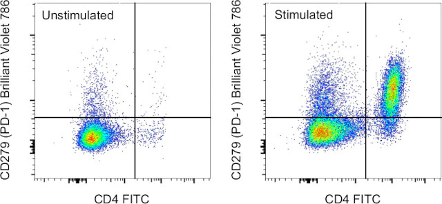
Description: The J43 monoclonal antibody reacts with mouse PD-1 (programmed death-1), a 55 kDa member of the Ig superfamily. PD-1 contains the immunoreceptor tyrosine-based inhibitory motif (ITIM) and plays a key role in peripheral tolerance and autoimmune disease in mice. PD-1 is expressed mainly on activated T and B lymphocytes. Two novel B7 Family members have been identified as PD-1 ligands, PD-L1 (B7-H1) and PD-L2 (B7-DC). Evidence reported to date suggests overlapping functions for these ligands and their constitutive expression on some normal tissues and upregulation on activated antigen-presenting cells. It is reported that J43 inhibits the binding of mouse PD-L1-Ig and mouse PD-L2-Ig to PD-1/BHK transfected cells. When administrated in vivo, both intact and Fab of J43 are reported to enhance contact hypersensitivity and exacerbate acute GVHD similar to transfer of PD-1-deficient cells. Injection of J43 also exacerbates EAE and NOD diabetes as do specific antibodies to mouse PD-L1 and PD-L2.
Applications Reported: This J43 antibody has been reported for use in flow cytometric analysis.
Applications Tested: This J43 antibody has been tested by flow cytometric analysis of mouse splenocytes. This may be used at less than or equal to 0.5 µg per test. A test is defined as the amount (µg) of antibody that will stain a cell sample in a final volume of 100 µL. Cell number should be determined empirically but can range from 10^5 to 10^8 cells/test. It is recommended that the antibody be carefully titrated for optimal performance in the assay of interest.
Brilliant Violet™ 786 (BV786) is a tandem dye that emits at 786 nm and is intended for use on cytometers equipped with a violet (405 nm) laser. Please make sure that your instrument is capable of detecting this fluorochrome.
When using two or more Super Bright, Brilliant Violet™, Brilliant Ultra Violet™, or other polymer dye-conjugated antibodies in a staining panel, it is recommended to use Super Bright Complete Staining Buffer (Product # SB-4401-42) or Brilliant Stain Buffer™ (Product # 00-4409-75) to minimize any non-specific polymer interactions. Please refer to the datasheet for Super Bright Staining Buffer or Brilliant Stain Buffer for more information.
Light sensitivity: This tandem dye is sensitive to photo-induced oxidation. Please protect this vial and stained samples from light.
Fixation: Samples can be stored in IC Fixation Buffer (Product # 00-8222-49) (100 µL of cell sample + 100 µL of IC Fixation Buffer) or 1-step Fix/Lyse Solution (Product # 00-5333-54) for up to 3 days in the dark at 4°C with minimal impact on brightness and FRET efficiency/compensation. Some generalizations regarding fluorophore performance after fixation can be made, but clone-specific performance should be determined empirically.
Our internal testing suggests that Brilliant Violet™ 786 (BV786) is not compatible with methanol-based fixation.
Excitation: 407 nm; Emission: 786 nm; Laser: Violet Laser.
BRILLIANT VIOLET™ is a trademark or registered trademark of Becton, Dickinson and Company or its affiliates, and is used under license. Powered by Sirigen™.
For Research Use Only. Not for use in diagnostic procedures. Not for resale without express authorization.
Original: $188.00
-70%$188.00
$56.40CD279 (PD-1) Monoclonal Antibody (J43), Brilliant Violet 786, eBioscience
PRODUCT DETAILS
Host: Armenian Hamster
Isotype: IgG
Clonality: Monoclonal
Clone: J43
Format: Brilliant Violet 786
Reactivity: Ms
Application: Flow Cytometry
Tested Dilution: 0.5 µg/test
Concentration: 0.2 mg/mL
Storage: 4°C, store in dark, DO NOT FREEZE!
Formulation: PBS with BSA and 0.09% sodium azide; pH 7.2
Purification: Affinity chromatography
Data Sheet: TDS
Specific Information
Description: The J43 monoclonal antibody reacts with mouse PD-1 (programmed death-1), a 55 kDa member of the Ig superfamily. PD-1 contains the immunoreceptor tyrosine-based inhibitory motif (ITIM) and plays a key role in peripheral tolerance and autoimmune disease in mice. PD-1 is expressed mainly on activated T and B lymphocytes. Two novel B7 Family members have been identified as PD-1 ligands, PD-L1 (B7-H1) and PD-L2 (B7-DC). Evidence reported to date suggests overlapping functions for these ligands and their constitutive expression on some normal tissues and upregulation on activated antigen-presenting cells. It is reported that J43 inhibits the binding of mouse PD-L1-Ig and mouse PD-L2-Ig to PD-1/BHK transfected cells. When administrated in vivo, both intact and Fab of J43 are reported to enhance contact hypersensitivity and exacerbate acute GVHD similar to transfer of PD-1-deficient cells. Injection of J43 also exacerbates EAE and NOD diabetes as do specific antibodies to mouse PD-L1 and PD-L2.
Applications Reported: This J43 antibody has been reported for use in flow cytometric analysis.
Applications Tested: This J43 antibody has been tested by flow cytometric analysis of mouse splenocytes. This may be used at less than or equal to 0.5 µg per test. A test is defined as the amount (µg) of antibody that will stain a cell sample in a final volume of 100 µL. Cell number should be determined empirically but can range from 10^5 to 10^8 cells/test. It is recommended that the antibody be carefully titrated for optimal performance in the assay of interest.
Brilliant Violet™ 786 (BV786) is a tandem dye that emits at 786 nm and is intended for use on cytometers equipped with a violet (405 nm) laser. Please make sure that your instrument is capable of detecting this fluorochrome.
When using two or more Super Bright, Brilliant Violet™, Brilliant Ultra Violet™, or other polymer dye-conjugated antibodies in a staining panel, it is recommended to use Super Bright Complete Staining Buffer (Product # SB-4401-42) or Brilliant Stain Buffer™ (Product # 00-4409-75) to minimize any non-specific polymer interactions. Please refer to the datasheet for Super Bright Staining Buffer or Brilliant Stain Buffer for more information.
Light sensitivity: This tandem dye is sensitive to photo-induced oxidation. Please protect this vial and stained samples from light.
Fixation: Samples can be stored in IC Fixation Buffer (Product # 00-8222-49) (100 µL of cell sample + 100 µL of IC Fixation Buffer) or 1-step Fix/Lyse Solution (Product # 00-5333-54) for up to 3 days in the dark at 4°C with minimal impact on brightness and FRET efficiency/compensation. Some generalizations regarding fluorophore performance after fixation can be made, but clone-specific performance should be determined empirically.
Our internal testing suggests that Brilliant Violet™ 786 (BV786) is not compatible with methanol-based fixation.
Excitation: 407 nm; Emission: 786 nm; Laser: Violet Laser.
BRILLIANT VIOLET™ is a trademark or registered trademark of Becton, Dickinson and Company or its affiliates, and is used under license. Powered by Sirigen™.
For Research Use Only. Not for use in diagnostic procedures. Not for resale without express authorization.
Product Information
Product Information
Shipping & Returns
Shipping & Returns
Description
PRODUCT DETAILS
Host: Armenian Hamster
Isotype: IgG
Clonality: Monoclonal
Clone: J43
Format: Brilliant Violet 786
Reactivity: Ms
Application: Flow Cytometry
Tested Dilution: 0.5 µg/test
Concentration: 0.2 mg/mL
Storage: 4°C, store in dark, DO NOT FREEZE!
Formulation: PBS with BSA and 0.09% sodium azide; pH 7.2
Purification: Affinity chromatography
Data Sheet: TDS
Specific Information
Description: The J43 monoclonal antibody reacts with mouse PD-1 (programmed death-1), a 55 kDa member of the Ig superfamily. PD-1 contains the immunoreceptor tyrosine-based inhibitory motif (ITIM) and plays a key role in peripheral tolerance and autoimmune disease in mice. PD-1 is expressed mainly on activated T and B lymphocytes. Two novel B7 Family members have been identified as PD-1 ligands, PD-L1 (B7-H1) and PD-L2 (B7-DC). Evidence reported to date suggests overlapping functions for these ligands and their constitutive expression on some normal tissues and upregulation on activated antigen-presenting cells. It is reported that J43 inhibits the binding of mouse PD-L1-Ig and mouse PD-L2-Ig to PD-1/BHK transfected cells. When administrated in vivo, both intact and Fab of J43 are reported to enhance contact hypersensitivity and exacerbate acute GVHD similar to transfer of PD-1-deficient cells. Injection of J43 also exacerbates EAE and NOD diabetes as do specific antibodies to mouse PD-L1 and PD-L2.
Applications Reported: This J43 antibody has been reported for use in flow cytometric analysis.
Applications Tested: This J43 antibody has been tested by flow cytometric analysis of mouse splenocytes. This may be used at less than or equal to 0.5 µg per test. A test is defined as the amount (µg) of antibody that will stain a cell sample in a final volume of 100 µL. Cell number should be determined empirically but can range from 10^5 to 10^8 cells/test. It is recommended that the antibody be carefully titrated for optimal performance in the assay of interest.
Brilliant Violet™ 786 (BV786) is a tandem dye that emits at 786 nm and is intended for use on cytometers equipped with a violet (405 nm) laser. Please make sure that your instrument is capable of detecting this fluorochrome.
When using two or more Super Bright, Brilliant Violet™, Brilliant Ultra Violet™, or other polymer dye-conjugated antibodies in a staining panel, it is recommended to use Super Bright Complete Staining Buffer (Product # SB-4401-42) or Brilliant Stain Buffer™ (Product # 00-4409-75) to minimize any non-specific polymer interactions. Please refer to the datasheet for Super Bright Staining Buffer or Brilliant Stain Buffer for more information.
Light sensitivity: This tandem dye is sensitive to photo-induced oxidation. Please protect this vial and stained samples from light.
Fixation: Samples can be stored in IC Fixation Buffer (Product # 00-8222-49) (100 µL of cell sample + 100 µL of IC Fixation Buffer) or 1-step Fix/Lyse Solution (Product # 00-5333-54) for up to 3 days in the dark at 4°C with minimal impact on brightness and FRET efficiency/compensation. Some generalizations regarding fluorophore performance after fixation can be made, but clone-specific performance should be determined empirically.
Our internal testing suggests that Brilliant Violet™ 786 (BV786) is not compatible with methanol-based fixation.
Excitation: 407 nm; Emission: 786 nm; Laser: Violet Laser.
BRILLIANT VIOLET™ is a trademark or registered trademark of Becton, Dickinson and Company or its affiliates, and is used under license. Powered by Sirigen™.
For Research Use Only. Not for use in diagnostic procedures. Not for resale without express authorization.


