CD272 (BTLA) Monoclonal Antibody (6G3), eBioscience
PRODUCT DETAILS
Host: Mouse
Isotype: IgG1, kappa
Clonality: Monoclonal
Clone: 6G3
Reactivity: Ms
Application: Flow Cytometry
Tested Dilution: 0.25 µg/test
Concentration: 0.5 mg/mL
Storage: 4°C
Formulation: PBS with 0.09% sodium azide; pH 7.2
Purification: Affinity chromatography
Data Sheet: TDS
Specific Information
Description: The 3F9.D12 antibody reacts with mouse BTLA, B and T lymphocyte attenuator from both BALB/c and C57Bl/6 strains. BTLA is expressed by peripheral lymphocytes, splenic macrophages, developing B cells in the bone marrow and developing T cells in thymus and mature, but not immature bone marrow-derived dendritic cells. BTLA is implicated as a negative regulator of the activation and/or function of various hemopoietic cell types. It is reported that BTLA binds to B7-H4, but further studies are needed to confirm this interaction.
Note: The anti-mouse BTLA monoclonal antibody 6F7 is reported to stain CD4+ and CD8+ single-positive (SP) thymocytes (Hurchla et al). However, other anti-mouse BTLA clones generated simultaneously with 6F7 (8F4, 3F9.D12, 6G3 and 6H6) do not stain SP thymocytes. It is not understood why there is a discrepancy in thymocyte staining however clones 8F4, 3F9.D12, 6G3 and 6H6 stain similar populations to 6F7 in splenocytes and bone marrow cells.
Applications Reported: This 6G3 antibody has been reported for use in flow cytometric analysis.
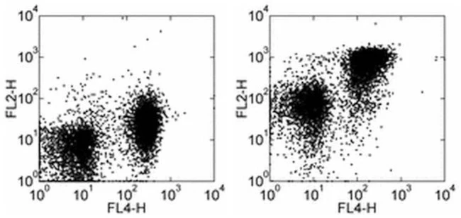
Applications Tested: This 6G3 antibody has been tested by flow cytometric analysis of mouse splenocytes. This can be used at less than or equal to 0.25 µg per test. A test is defined as the amount (µg) of antibody that will stain a cell sample in a final volume of 100 µL. Cell number should be determined empirically but can range from 10^5 to 10^8 cells/test. It is recommended that the antibody be carefully titrated for optimal performance in the assay of interest.
Purity: Greater than 90%, as determined by SDS-PAGE.
Aggregation: Less than 10%, as determined by HPLC.
Filtration: 0.2 µm post-manufacturing filtered.
For Research Use Only. Not for use in diagnostic procedures. Not for resale without express authorization.
Original: $366.00
-70%$366.00
$109.80CD272 (BTLA) Monoclonal Antibody (6G3), eBioscience
PRODUCT DETAILS
Host: Mouse
Isotype: IgG1, kappa
Clonality: Monoclonal
Clone: 6G3
Reactivity: Ms
Application: Flow Cytometry
Tested Dilution: 0.25 µg/test
Concentration: 0.5 mg/mL
Storage: 4°C
Formulation: PBS with 0.09% sodium azide; pH 7.2
Purification: Affinity chromatography
Data Sheet: TDS
Specific Information
Description: The 3F9.D12 antibody reacts with mouse BTLA, B and T lymphocyte attenuator from both BALB/c and C57Bl/6 strains. BTLA is expressed by peripheral lymphocytes, splenic macrophages, developing B cells in the bone marrow and developing T cells in thymus and mature, but not immature bone marrow-derived dendritic cells. BTLA is implicated as a negative regulator of the activation and/or function of various hemopoietic cell types. It is reported that BTLA binds to B7-H4, but further studies are needed to confirm this interaction.
Note: The anti-mouse BTLA monoclonal antibody 6F7 is reported to stain CD4+ and CD8+ single-positive (SP) thymocytes (Hurchla et al). However, other anti-mouse BTLA clones generated simultaneously with 6F7 (8F4, 3F9.D12, 6G3 and 6H6) do not stain SP thymocytes. It is not understood why there is a discrepancy in thymocyte staining however clones 8F4, 3F9.D12, 6G3 and 6H6 stain similar populations to 6F7 in splenocytes and bone marrow cells.
Applications Reported: This 6G3 antibody has been reported for use in flow cytometric analysis.
Applications Tested: This 6G3 antibody has been tested by flow cytometric analysis of mouse splenocytes. This can be used at less than or equal to 0.25 µg per test. A test is defined as the amount (µg) of antibody that will stain a cell sample in a final volume of 100 µL. Cell number should be determined empirically but can range from 10^5 to 10^8 cells/test. It is recommended that the antibody be carefully titrated for optimal performance in the assay of interest.
Purity: Greater than 90%, as determined by SDS-PAGE.
Aggregation: Less than 10%, as determined by HPLC.
Filtration: 0.2 µm post-manufacturing filtered.
For Research Use Only. Not for use in diagnostic procedures. Not for resale without express authorization.
Product Information
Product Information
Shipping & Returns
Shipping & Returns
Description
PRODUCT DETAILS
Host: Mouse
Isotype: IgG1, kappa
Clonality: Monoclonal
Clone: 6G3
Reactivity: Ms
Application: Flow Cytometry
Tested Dilution: 0.25 µg/test
Concentration: 0.5 mg/mL
Storage: 4°C
Formulation: PBS with 0.09% sodium azide; pH 7.2
Purification: Affinity chromatography
Data Sheet: TDS
Specific Information
Description: The 3F9.D12 antibody reacts with mouse BTLA, B and T lymphocyte attenuator from both BALB/c and C57Bl/6 strains. BTLA is expressed by peripheral lymphocytes, splenic macrophages, developing B cells in the bone marrow and developing T cells in thymus and mature, but not immature bone marrow-derived dendritic cells. BTLA is implicated as a negative regulator of the activation and/or function of various hemopoietic cell types. It is reported that BTLA binds to B7-H4, but further studies are needed to confirm this interaction.
Note: The anti-mouse BTLA monoclonal antibody 6F7 is reported to stain CD4+ and CD8+ single-positive (SP) thymocytes (Hurchla et al). However, other anti-mouse BTLA clones generated simultaneously with 6F7 (8F4, 3F9.D12, 6G3 and 6H6) do not stain SP thymocytes. It is not understood why there is a discrepancy in thymocyte staining however clones 8F4, 3F9.D12, 6G3 and 6H6 stain similar populations to 6F7 in splenocytes and bone marrow cells.
Applications Reported: This 6G3 antibody has been reported for use in flow cytometric analysis.
Applications Tested: This 6G3 antibody has been tested by flow cytometric analysis of mouse splenocytes. This can be used at less than or equal to 0.25 µg per test. A test is defined as the amount (µg) of antibody that will stain a cell sample in a final volume of 100 µL. Cell number should be determined empirically but can range from 10^5 to 10^8 cells/test. It is recommended that the antibody be carefully titrated for optimal performance in the assay of interest.
Purity: Greater than 90%, as determined by SDS-PAGE.
Aggregation: Less than 10%, as determined by HPLC.
Filtration: 0.2 µm post-manufacturing filtered.
For Research Use Only. Not for use in diagnostic procedures. Not for resale without express authorization.


