CD206 (MMR) Monoclonal Antibody (MR6F3), Alexa Fluor 488, eBioscience
PRODUCT DETAILS
Host: Rat
Isotype: IgG2b, kappa
Clonality: Monoclonal
Clone: MR6F3
Format: Alexa Fluor 488
Reactivity: Ms
Application: Flow Cytometry
Tested Dilution: 1.0 µg/test
Concentration: 0.5 mg/mL
Storage: 4°C, store in dark, DO NOT FREEZE!
Formulation: PBS with 0.09% sodium azide; pH 7.2
Purification: Affinity chromatography
Data Sheet: TDS
Specific Information
Description: This MR6F3 antibody recognizes mouse CD206 also known as Macrophage Mannose Receptor (MMR) or Mannose Receptor C, Type 1 (MRC1). CD206 is a 175-kDa, type 1 integral membrane glycoprotein receptor that is present in macrophages, some dendritic cells, as well as liver and lymphoid endothelial cells. CD206 belongs to the C-type lectin family. Extracellular regions of CD206 include an N-terminal cysteine-rich (CR) domain that binds sulfated glycoproteins, a fibronectin II (FNII) domain that binds collagens, and eight carbohydrate recognition domains (CRDs) that bind sugars like mannose and fucose with high affinity. CD206 mediates phagocytic and endocytic uptake of fungal, bacterial, protozoan and viral antigens, and plays an important role in immune defense and immune regulation. A soluble form of CD206 is generated by cleavage of the full-length protein, and it can be detected in in vitro macrophage cell culture supernatants and in mouse serum. CD206 is considered to be one of the markers of M2 macrophages. Factors inducing its expression include: IL-4, IL-13, M-CSF, IL-6, IL-10, and glucocorticoids, while TNF-alpha, IFN-gamma, TGF-beta, and LPS have been reported to down-regulate expression of CD206.
It has been demonstrated that only a small fraction of CD206 is present at the cell surface, therefore, intracellular staining is recommended.
Applications Reported: This MR6F3 antibody has been reported for use in flow cytometric analysis.
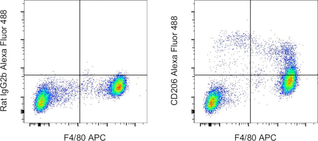
Applications Tested: This MR6F3 antibody has been tested by flow cytometric analysis of resident peritoneal macrophages. This may be used at less than or equal to 1 µg per test. A test is defined as the amount (µg) of antibody that will stain a cell sample in a final volume of 100 µL. Cell number should be determined empirically but can range from 10^5 to 10^8 cells/test. It is recommended that the antibody be carefully titrated for optimal performance in the assay of interest.
Excitation: 488 nm; Emission: 519 nm; Laser: Blue Laser"
For Research Use Only. Not for use in diagnostic procedures. Not for resale without express authorization.
Original: $370.00
-70%$370.00
$111.00CD206 (MMR) Monoclonal Antibody (MR6F3), Alexa Fluor 488, eBioscience
PRODUCT DETAILS
Host: Rat
Isotype: IgG2b, kappa
Clonality: Monoclonal
Clone: MR6F3
Format: Alexa Fluor 488
Reactivity: Ms
Application: Flow Cytometry
Tested Dilution: 1.0 µg/test
Concentration: 0.5 mg/mL
Storage: 4°C, store in dark, DO NOT FREEZE!
Formulation: PBS with 0.09% sodium azide; pH 7.2
Purification: Affinity chromatography
Data Sheet: TDS
Specific Information
Description: This MR6F3 antibody recognizes mouse CD206 also known as Macrophage Mannose Receptor (MMR) or Mannose Receptor C, Type 1 (MRC1). CD206 is a 175-kDa, type 1 integral membrane glycoprotein receptor that is present in macrophages, some dendritic cells, as well as liver and lymphoid endothelial cells. CD206 belongs to the C-type lectin family. Extracellular regions of CD206 include an N-terminal cysteine-rich (CR) domain that binds sulfated glycoproteins, a fibronectin II (FNII) domain that binds collagens, and eight carbohydrate recognition domains (CRDs) that bind sugars like mannose and fucose with high affinity. CD206 mediates phagocytic and endocytic uptake of fungal, bacterial, protozoan and viral antigens, and plays an important role in immune defense and immune regulation. A soluble form of CD206 is generated by cleavage of the full-length protein, and it can be detected in in vitro macrophage cell culture supernatants and in mouse serum. CD206 is considered to be one of the markers of M2 macrophages. Factors inducing its expression include: IL-4, IL-13, M-CSF, IL-6, IL-10, and glucocorticoids, while TNF-alpha, IFN-gamma, TGF-beta, and LPS have been reported to down-regulate expression of CD206.
It has been demonstrated that only a small fraction of CD206 is present at the cell surface, therefore, intracellular staining is recommended.
Applications Reported: This MR6F3 antibody has been reported for use in flow cytometric analysis.
Applications Tested: This MR6F3 antibody has been tested by flow cytometric analysis of resident peritoneal macrophages. This may be used at less than or equal to 1 µg per test. A test is defined as the amount (µg) of antibody that will stain a cell sample in a final volume of 100 µL. Cell number should be determined empirically but can range from 10^5 to 10^8 cells/test. It is recommended that the antibody be carefully titrated for optimal performance in the assay of interest.
Excitation: 488 nm; Emission: 519 nm; Laser: Blue Laser"
For Research Use Only. Not for use in diagnostic procedures. Not for resale without express authorization.
Product Information
Product Information
Shipping & Returns
Shipping & Returns
Description
PRODUCT DETAILS
Host: Rat
Isotype: IgG2b, kappa
Clonality: Monoclonal
Clone: MR6F3
Format: Alexa Fluor 488
Reactivity: Ms
Application: Flow Cytometry
Tested Dilution: 1.0 µg/test
Concentration: 0.5 mg/mL
Storage: 4°C, store in dark, DO NOT FREEZE!
Formulation: PBS with 0.09% sodium azide; pH 7.2
Purification: Affinity chromatography
Data Sheet: TDS
Specific Information
Description: This MR6F3 antibody recognizes mouse CD206 also known as Macrophage Mannose Receptor (MMR) or Mannose Receptor C, Type 1 (MRC1). CD206 is a 175-kDa, type 1 integral membrane glycoprotein receptor that is present in macrophages, some dendritic cells, as well as liver and lymphoid endothelial cells. CD206 belongs to the C-type lectin family. Extracellular regions of CD206 include an N-terminal cysteine-rich (CR) domain that binds sulfated glycoproteins, a fibronectin II (FNII) domain that binds collagens, and eight carbohydrate recognition domains (CRDs) that bind sugars like mannose and fucose with high affinity. CD206 mediates phagocytic and endocytic uptake of fungal, bacterial, protozoan and viral antigens, and plays an important role in immune defense and immune regulation. A soluble form of CD206 is generated by cleavage of the full-length protein, and it can be detected in in vitro macrophage cell culture supernatants and in mouse serum. CD206 is considered to be one of the markers of M2 macrophages. Factors inducing its expression include: IL-4, IL-13, M-CSF, IL-6, IL-10, and glucocorticoids, while TNF-alpha, IFN-gamma, TGF-beta, and LPS have been reported to down-regulate expression of CD206.
It has been demonstrated that only a small fraction of CD206 is present at the cell surface, therefore, intracellular staining is recommended.
Applications Reported: This MR6F3 antibody has been reported for use in flow cytometric analysis.
Applications Tested: This MR6F3 antibody has been tested by flow cytometric analysis of resident peritoneal macrophages. This may be used at less than or equal to 1 µg per test. A test is defined as the amount (µg) of antibody that will stain a cell sample in a final volume of 100 µL. Cell number should be determined empirically but can range from 10^5 to 10^8 cells/test. It is recommended that the antibody be carefully titrated for optimal performance in the assay of interest.
Excitation: 488 nm; Emission: 519 nm; Laser: Blue Laser"
For Research Use Only. Not for use in diagnostic procedures. Not for resale without express authorization.


