CD206 (MMR) Monoclonal Antibody (19.2), PE-Cyanine7, eBioscience
PRODUCT DETAILS
Host: Mouse
Isotype: IgG1, kappa
Clonality: Monoclonal
Clone: 19.2
Format: PE-Cyanine7
Reactivity: Hu
Application: Flow Cytometry
Tested Dilution: 5 µL (0.06 µg)/test
Concentration: 5 μL/Test
Storage: 4°C, store in dark, DO NOT FREEZE!
Formulation: PBS with BSA and 0.09% sodium azide; pH 7.2
Purification: Affinity chromatography
Data Sheet: TDS
Specific Information
Description: This 19.2 monoclonal antibody reacts with human CD206, which is also known as the macrophage mannose receptor (MMR). CD206 is expressed on macrophages and dendritic cells. This type I transmembrane protein can also be detected on non-immune cells, including hepatic and lymphathic epithelia and kidney mesengial cells. CD206 binds to glycoproteins that terminate in D-mannose, L-fucose, or N-acetylglucosamine, as well as a variety of hormones. This receptor undergoes constitutive internalization and recycling between the plasma membrane and the endosomal compartment. CD206 is involved in antigen processing and presentation, cell migration, and intracellular signaling. Moreover, CD206 plays a key role in phagocytosis pathogens such as Candida albicans, Leishmania, and Mycobacterium tuberculosis.
Applications Reported: This 19.2 antibody has been reported for use in flow cytometric analysis.
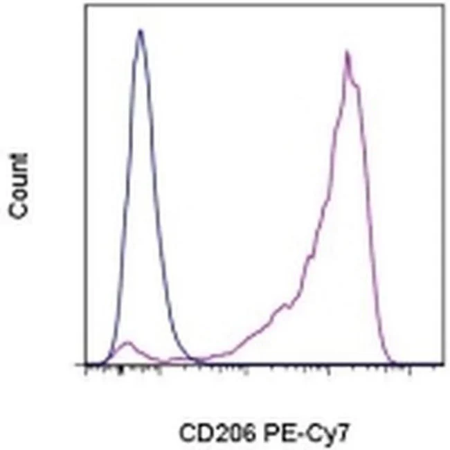
Applications Tested: This 19.2 antibody has been pre-titrated and tested by flow cytometric analysis of stimulated human monocytes. This can be used at 5 µL (0.06 µg) per test. A test is defined as the amount (µg) of antibody that will stain a cell sample in a final volume of 100 µL. Cell number should be determined empirically but can range from 10^5 to 10^8 cells/test.
Light sensitivity: This tandem dye is sensitive photo-induced oxidation. Please protect this vial and stained samples from light.
Fixation: Samples can be stored in IC Fixation Buffer (Product # 00-822-49) (100 µL cell sample + 100 µL IC Fixation Buffer) or 1-step Fix/Lyse Solution (Product # 00-5333-54) for up to 3 days in the dark at 4°C with minimal impact on brightness and FRET efficiency/compensation. Some generalizations regarding fluorophore performance after fixation can be made, but clone specific performance should be determined empirically.
Excitation: 488-561 nm; Emission: 775 nm; Laser: Blue Laser, Green Laser, Yellow-Green Laser.
Filtration: 0.2 µm post-manufacturing filtered.
For Research Use Only. Not for use in diagnostic procedures. Not for resale without express authorization.
Original: $436.00
-70%$436.00
$130.80CD206 (MMR) Monoclonal Antibody (19.2), PE-Cyanine7, eBioscience
PRODUCT DETAILS
Host: Mouse
Isotype: IgG1, kappa
Clonality: Monoclonal
Clone: 19.2
Format: PE-Cyanine7
Reactivity: Hu
Application: Flow Cytometry
Tested Dilution: 5 µL (0.06 µg)/test
Concentration: 5 μL/Test
Storage: 4°C, store in dark, DO NOT FREEZE!
Formulation: PBS with BSA and 0.09% sodium azide; pH 7.2
Purification: Affinity chromatography
Data Sheet: TDS
Specific Information
Description: This 19.2 monoclonal antibody reacts with human CD206, which is also known as the macrophage mannose receptor (MMR). CD206 is expressed on macrophages and dendritic cells. This type I transmembrane protein can also be detected on non-immune cells, including hepatic and lymphathic epithelia and kidney mesengial cells. CD206 binds to glycoproteins that terminate in D-mannose, L-fucose, or N-acetylglucosamine, as well as a variety of hormones. This receptor undergoes constitutive internalization and recycling between the plasma membrane and the endosomal compartment. CD206 is involved in antigen processing and presentation, cell migration, and intracellular signaling. Moreover, CD206 plays a key role in phagocytosis pathogens such as Candida albicans, Leishmania, and Mycobacterium tuberculosis.
Applications Reported: This 19.2 antibody has been reported for use in flow cytometric analysis.
Applications Tested: This 19.2 antibody has been pre-titrated and tested by flow cytometric analysis of stimulated human monocytes. This can be used at 5 µL (0.06 µg) per test. A test is defined as the amount (µg) of antibody that will stain a cell sample in a final volume of 100 µL. Cell number should be determined empirically but can range from 10^5 to 10^8 cells/test.
Light sensitivity: This tandem dye is sensitive photo-induced oxidation. Please protect this vial and stained samples from light.
Fixation: Samples can be stored in IC Fixation Buffer (Product # 00-822-49) (100 µL cell sample + 100 µL IC Fixation Buffer) or 1-step Fix/Lyse Solution (Product # 00-5333-54) for up to 3 days in the dark at 4°C with minimal impact on brightness and FRET efficiency/compensation. Some generalizations regarding fluorophore performance after fixation can be made, but clone specific performance should be determined empirically.
Excitation: 488-561 nm; Emission: 775 nm; Laser: Blue Laser, Green Laser, Yellow-Green Laser.
Filtration: 0.2 µm post-manufacturing filtered.
For Research Use Only. Not for use in diagnostic procedures. Not for resale without express authorization.
Product Information
Product Information
Shipping & Returns
Shipping & Returns
Description
PRODUCT DETAILS
Host: Mouse
Isotype: IgG1, kappa
Clonality: Monoclonal
Clone: 19.2
Format: PE-Cyanine7
Reactivity: Hu
Application: Flow Cytometry
Tested Dilution: 5 µL (0.06 µg)/test
Concentration: 5 μL/Test
Storage: 4°C, store in dark, DO NOT FREEZE!
Formulation: PBS with BSA and 0.09% sodium azide; pH 7.2
Purification: Affinity chromatography
Data Sheet: TDS
Specific Information
Description: This 19.2 monoclonal antibody reacts with human CD206, which is also known as the macrophage mannose receptor (MMR). CD206 is expressed on macrophages and dendritic cells. This type I transmembrane protein can also be detected on non-immune cells, including hepatic and lymphathic epithelia and kidney mesengial cells. CD206 binds to glycoproteins that terminate in D-mannose, L-fucose, or N-acetylglucosamine, as well as a variety of hormones. This receptor undergoes constitutive internalization and recycling between the plasma membrane and the endosomal compartment. CD206 is involved in antigen processing and presentation, cell migration, and intracellular signaling. Moreover, CD206 plays a key role in phagocytosis pathogens such as Candida albicans, Leishmania, and Mycobacterium tuberculosis.
Applications Reported: This 19.2 antibody has been reported for use in flow cytometric analysis.
Applications Tested: This 19.2 antibody has been pre-titrated and tested by flow cytometric analysis of stimulated human monocytes. This can be used at 5 µL (0.06 µg) per test. A test is defined as the amount (µg) of antibody that will stain a cell sample in a final volume of 100 µL. Cell number should be determined empirically but can range from 10^5 to 10^8 cells/test.
Light sensitivity: This tandem dye is sensitive photo-induced oxidation. Please protect this vial and stained samples from light.
Fixation: Samples can be stored in IC Fixation Buffer (Product # 00-822-49) (100 µL cell sample + 100 µL IC Fixation Buffer) or 1-step Fix/Lyse Solution (Product # 00-5333-54) for up to 3 days in the dark at 4°C with minimal impact on brightness and FRET efficiency/compensation. Some generalizations regarding fluorophore performance after fixation can be made, but clone specific performance should be determined empirically.
Excitation: 488-561 nm; Emission: 775 nm; Laser: Blue Laser, Green Laser, Yellow-Green Laser.
Filtration: 0.2 µm post-manufacturing filtered.
For Research Use Only. Not for use in diagnostic procedures. Not for resale without express authorization.


