CD1c Monoclonal Antibody (L161), Super Bright 436, eBioscience
PRODUCT DETAILS
Host: Mouse
Isotype: IgG1, kappa
Clonality: Monoclonal
Clone: L161
Format: Super Bright 436
Reactivity: Hu
Application: Flow Cytometry
Tested Dilution: 5 µL (0.25 µg) /test
Concentration: 5 μL/Test
Storage: 4°C, store in dark, DO NOT FREEZE!
Formulation: PBS with BSA and 0.09% sodium azide; pH 7.2
Purification: Affinity chromatography
Data Sheet: TDS
Specific Information
Description: This L161 monoclonal antibody detects CD1c (also known as BDCA-1), a glycoprotein that is noncovalently linked to beta-2 microglobulin on thymocytes and antigen presenting cells such as dendritic and Langerhans cells. This molecule is also expressed on some circulating and marginal zone B cells, as well as in lymph nodes and germinal centers. CD1c is involved in the presentation of lipid antigens such as microbial fatty acids to effector T cells during the adaptive immune response. Finally, alternative splicing gives rise to three different isoforms of CD1c (soluble, membrane, and cytoplasmic/soluble isoforms).
Applications Reported: This L161 antibody has been reported for use in flow cytometric analysis.
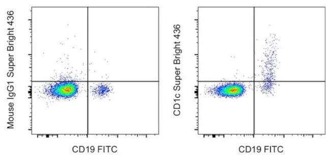
Applications Tested: This L161 antibody has been pre-diluted and tested by flow cytometric analysis of normal human peripheral blood cells. This may be used at 5 µL (0.25 µg) per test. A test is defined as the amount (µg) of antibody that will stain a cell sample in a final volume of 100 µL. Cell number should be determined empirically but can range from 10^5 to 10^8 cells/test.
Super Bright 436 can be excited with the violet laser line (405 nm) and emits at 436 nm. We recommend using a 450/50 bandpass filter, or equivalent. Please make sure that your instrument is capable of detecting this fluorochrome.
When using two or more Super Bright dye-conjugated antibodies in a staining panel, it is recommended to use Super Bright Complete Staining Buffer (Product # SB-4401) to minimize any non-specific polymer interactions. Please refer to the datasheet for Super Bright Staining Buffer for more information.
Excitation: 405 nm; Emission: 436 nm; Laser: Violet Laser
Super Bright Polymer Dyes are sold under license from Becton, Dickinson and Company.
For Research Use Only. Not for use in diagnostic procedures. Not for resale without express authorization.
Original: $380.00
-70%$380.00
$114.00CD1c Monoclonal Antibody (L161), Super Bright 436, eBioscience
PRODUCT DETAILS
Host: Mouse
Isotype: IgG1, kappa
Clonality: Monoclonal
Clone: L161
Format: Super Bright 436
Reactivity: Hu
Application: Flow Cytometry
Tested Dilution: 5 µL (0.25 µg) /test
Concentration: 5 μL/Test
Storage: 4°C, store in dark, DO NOT FREEZE!
Formulation: PBS with BSA and 0.09% sodium azide; pH 7.2
Purification: Affinity chromatography
Data Sheet: TDS
Specific Information
Description: This L161 monoclonal antibody detects CD1c (also known as BDCA-1), a glycoprotein that is noncovalently linked to beta-2 microglobulin on thymocytes and antigen presenting cells such as dendritic and Langerhans cells. This molecule is also expressed on some circulating and marginal zone B cells, as well as in lymph nodes and germinal centers. CD1c is involved in the presentation of lipid antigens such as microbial fatty acids to effector T cells during the adaptive immune response. Finally, alternative splicing gives rise to three different isoforms of CD1c (soluble, membrane, and cytoplasmic/soluble isoforms).
Applications Reported: This L161 antibody has been reported for use in flow cytometric analysis.
Applications Tested: This L161 antibody has been pre-diluted and tested by flow cytometric analysis of normal human peripheral blood cells. This may be used at 5 µL (0.25 µg) per test. A test is defined as the amount (µg) of antibody that will stain a cell sample in a final volume of 100 µL. Cell number should be determined empirically but can range from 10^5 to 10^8 cells/test.
Super Bright 436 can be excited with the violet laser line (405 nm) and emits at 436 nm. We recommend using a 450/50 bandpass filter, or equivalent. Please make sure that your instrument is capable of detecting this fluorochrome.
When using two or more Super Bright dye-conjugated antibodies in a staining panel, it is recommended to use Super Bright Complete Staining Buffer (Product # SB-4401) to minimize any non-specific polymer interactions. Please refer to the datasheet for Super Bright Staining Buffer for more information.
Excitation: 405 nm; Emission: 436 nm; Laser: Violet Laser
Super Bright Polymer Dyes are sold under license from Becton, Dickinson and Company.
For Research Use Only. Not for use in diagnostic procedures. Not for resale without express authorization.
Product Information
Product Information
Shipping & Returns
Shipping & Returns
Description
PRODUCT DETAILS
Host: Mouse
Isotype: IgG1, kappa
Clonality: Monoclonal
Clone: L161
Format: Super Bright 436
Reactivity: Hu
Application: Flow Cytometry
Tested Dilution: 5 µL (0.25 µg) /test
Concentration: 5 μL/Test
Storage: 4°C, store in dark, DO NOT FREEZE!
Formulation: PBS with BSA and 0.09% sodium azide; pH 7.2
Purification: Affinity chromatography
Data Sheet: TDS
Specific Information
Description: This L161 monoclonal antibody detects CD1c (also known as BDCA-1), a glycoprotein that is noncovalently linked to beta-2 microglobulin on thymocytes and antigen presenting cells such as dendritic and Langerhans cells. This molecule is also expressed on some circulating and marginal zone B cells, as well as in lymph nodes and germinal centers. CD1c is involved in the presentation of lipid antigens such as microbial fatty acids to effector T cells during the adaptive immune response. Finally, alternative splicing gives rise to three different isoforms of CD1c (soluble, membrane, and cytoplasmic/soluble isoforms).
Applications Reported: This L161 antibody has been reported for use in flow cytometric analysis.
Applications Tested: This L161 antibody has been pre-diluted and tested by flow cytometric analysis of normal human peripheral blood cells. This may be used at 5 µL (0.25 µg) per test. A test is defined as the amount (µg) of antibody that will stain a cell sample in a final volume of 100 µL. Cell number should be determined empirically but can range from 10^5 to 10^8 cells/test.
Super Bright 436 can be excited with the violet laser line (405 nm) and emits at 436 nm. We recommend using a 450/50 bandpass filter, or equivalent. Please make sure that your instrument is capable of detecting this fluorochrome.
When using two or more Super Bright dye-conjugated antibodies in a staining panel, it is recommended to use Super Bright Complete Staining Buffer (Product # SB-4401) to minimize any non-specific polymer interactions. Please refer to the datasheet for Super Bright Staining Buffer for more information.
Excitation: 405 nm; Emission: 436 nm; Laser: Violet Laser
Super Bright Polymer Dyes are sold under license from Becton, Dickinson and Company.
For Research Use Only. Not for use in diagnostic procedures. Not for resale without express authorization.


