CD172a (SIRP alpha) Monoclonal Antibody (P84), Super Bright 436, eBioscience
PRODUCT DETAILS
Host: Rat
Isotype: IgG1, kappa
Clonality: Monoclonal
Clone: P84
Format: Super Bright 436
Reactivity: Ms
Application: Flow Cytometry
Tested Dilution: 1.0 µg/test
Concentration: 0.2 mg/mL
Storage: 4°C, store in dark, DO NOT FREEZE!
Formulation: PBS with BSA and 0.09% sodium azide; pH 7.2
Purification: Affinity chromatography
Data Sheet: TDS
Specific Information
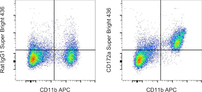
Description: This P84 monoclonal antibody reacts with mouse CD172a, also known as signal regulatory protein a (SIRPa). This cell surface glycoprotein consists of three Ig-like extracellular domains and two cytoplasmic immunoreceptor tyrosine-based inhibitory motifs (ITIMs). The ITIM domains have been demonstrated to recruit and bind the Src homology 2 domain-containing phosphatases SHP-1 and SHP-2. CD172a is expressed on monocytes, macrophages, dendritic cells, but not on T and B lymphocytes. Moreover, neurons and other tissues of the central nervous system have also been shown to express CD172a. The integrin-associated protein CD47 is the extracellular ligand for CD172a. Studies show that CD172a is involved in dendritic cell-mediated T cell activation, neutrophil migration, and phagocytosis.
This monoclonal antibody has been reported to have neutralizing activity.
Applications Reported: This P84 antibody has been reported for use in flow cytometric analysis.
Applications Tested: This P84 antibody has been tested by flow cytometric analysis of mouse bone marrow cells. This may be used at less than or equal to 1.0 µg per test. A test is defined as the amount (µg) of antibody that will stain a cell sample in a final volume of 100 µL. Cell number should be determined empirically but can range from 10^5 to 10^8 cells/test. It is recommended that the antibody be carefully titrated for optimal performance in the assay of interest.
Super Bright 436 can be excited with the violet laser line (405 nm) and emits at 436 nm. We recommend using a 450/50 bandpass filter, or equivalent. Please make sure that your instrument is capable of detecting this fluorochrome.
When using two or more Super Bright dye-conjugated antibodies in a staining panel, it is recommended to use Super Bright Complete Staining Buffer (Product # SB-4401) to minimize any non-specific polymer interactions. Please refer to the datasheet for Super Bright Staining Buffer for more information.
Excitation: 405 nm; Emission: 436 nm; Laser: Violet Laser
Super Bright Polymer Dyes are sold under license from Becton, Dickinson and Company.
For Research Use Only. Not for use in diagnostic procedures. Not for resale without express authorization.
Original: $186.00
-70%$186.00
$55.80CD172a (SIRP alpha) Monoclonal Antibody (P84), Super Bright 436, eBioscience
PRODUCT DETAILS
Host: Rat
Isotype: IgG1, kappa
Clonality: Monoclonal
Clone: P84
Format: Super Bright 436
Reactivity: Ms
Application: Flow Cytometry
Tested Dilution: 1.0 µg/test
Concentration: 0.2 mg/mL
Storage: 4°C, store in dark, DO NOT FREEZE!
Formulation: PBS with BSA and 0.09% sodium azide; pH 7.2
Purification: Affinity chromatography
Data Sheet: TDS
Specific Information
Description: This P84 monoclonal antibody reacts with mouse CD172a, also known as signal regulatory protein a (SIRPa). This cell surface glycoprotein consists of three Ig-like extracellular domains and two cytoplasmic immunoreceptor tyrosine-based inhibitory motifs (ITIMs). The ITIM domains have been demonstrated to recruit and bind the Src homology 2 domain-containing phosphatases SHP-1 and SHP-2. CD172a is expressed on monocytes, macrophages, dendritic cells, but not on T and B lymphocytes. Moreover, neurons and other tissues of the central nervous system have also been shown to express CD172a. The integrin-associated protein CD47 is the extracellular ligand for CD172a. Studies show that CD172a is involved in dendritic cell-mediated T cell activation, neutrophil migration, and phagocytosis.
This monoclonal antibody has been reported to have neutralizing activity.
Applications Reported: This P84 antibody has been reported for use in flow cytometric analysis.
Applications Tested: This P84 antibody has been tested by flow cytometric analysis of mouse bone marrow cells. This may be used at less than or equal to 1.0 µg per test. A test is defined as the amount (µg) of antibody that will stain a cell sample in a final volume of 100 µL. Cell number should be determined empirically but can range from 10^5 to 10^8 cells/test. It is recommended that the antibody be carefully titrated for optimal performance in the assay of interest.
Super Bright 436 can be excited with the violet laser line (405 nm) and emits at 436 nm. We recommend using a 450/50 bandpass filter, or equivalent. Please make sure that your instrument is capable of detecting this fluorochrome.
When using two or more Super Bright dye-conjugated antibodies in a staining panel, it is recommended to use Super Bright Complete Staining Buffer (Product # SB-4401) to minimize any non-specific polymer interactions. Please refer to the datasheet for Super Bright Staining Buffer for more information.
Excitation: 405 nm; Emission: 436 nm; Laser: Violet Laser
Super Bright Polymer Dyes are sold under license from Becton, Dickinson and Company.
For Research Use Only. Not for use in diagnostic procedures. Not for resale without express authorization.
Product Information
Product Information
Shipping & Returns
Shipping & Returns
Description
PRODUCT DETAILS
Host: Rat
Isotype: IgG1, kappa
Clonality: Monoclonal
Clone: P84
Format: Super Bright 436
Reactivity: Ms
Application: Flow Cytometry
Tested Dilution: 1.0 µg/test
Concentration: 0.2 mg/mL
Storage: 4°C, store in dark, DO NOT FREEZE!
Formulation: PBS with BSA and 0.09% sodium azide; pH 7.2
Purification: Affinity chromatography
Data Sheet: TDS
Specific Information
Description: This P84 monoclonal antibody reacts with mouse CD172a, also known as signal regulatory protein a (SIRPa). This cell surface glycoprotein consists of three Ig-like extracellular domains and two cytoplasmic immunoreceptor tyrosine-based inhibitory motifs (ITIMs). The ITIM domains have been demonstrated to recruit and bind the Src homology 2 domain-containing phosphatases SHP-1 and SHP-2. CD172a is expressed on monocytes, macrophages, dendritic cells, but not on T and B lymphocytes. Moreover, neurons and other tissues of the central nervous system have also been shown to express CD172a. The integrin-associated protein CD47 is the extracellular ligand for CD172a. Studies show that CD172a is involved in dendritic cell-mediated T cell activation, neutrophil migration, and phagocytosis.
This monoclonal antibody has been reported to have neutralizing activity.
Applications Reported: This P84 antibody has been reported for use in flow cytometric analysis.
Applications Tested: This P84 antibody has been tested by flow cytometric analysis of mouse bone marrow cells. This may be used at less than or equal to 1.0 µg per test. A test is defined as the amount (µg) of antibody that will stain a cell sample in a final volume of 100 µL. Cell number should be determined empirically but can range from 10^5 to 10^8 cells/test. It is recommended that the antibody be carefully titrated for optimal performance in the assay of interest.
Super Bright 436 can be excited with the violet laser line (405 nm) and emits at 436 nm. We recommend using a 450/50 bandpass filter, or equivalent. Please make sure that your instrument is capable of detecting this fluorochrome.
When using two or more Super Bright dye-conjugated antibodies in a staining panel, it is recommended to use Super Bright Complete Staining Buffer (Product # SB-4401) to minimize any non-specific polymer interactions. Please refer to the datasheet for Super Bright Staining Buffer for more information.
Excitation: 405 nm; Emission: 436 nm; Laser: Violet Laser
Super Bright Polymer Dyes are sold under license from Becton, Dickinson and Company.
For Research Use Only. Not for use in diagnostic procedures. Not for resale without express authorization.


