CD146 Monoclonal Antibody (P1H12), NovaFluor Blue 610-70S, eBioscience
PRODUCT DETAILS
Host: Mouse
Isotype: IgG1, kappa
Clonality: Monoclonal
Clone: P1H12
Format: NovaFluor Blue 610-70S
Reactivity: Ca, Hu, Ms, Rb
Application: Flow Cytometry
Tested Dilution: 5 µL (0.2 µg)/test
Concentration: 0.2 μg/Test
Storage: 4°C, store in dark, DO NOT FREEZE!
Formulation: PBS with 0.09% sodium azide; pH 7.2
Purification: Affinity chromatography
Data Sheet: TDS
Specific Information
Description: The monoclonal antibody P1H12 recognizes CD146 also known as MUC18, s-endo, Endo-CAM and Mel-CAM, which is a member of the Ig superfamily of proteins. The expression of CD146 is found on endothelial cells, bone marrow fibroblasts and some tumors (especially melanoma). Recently mesenchymal stromal cells and endometrial stromal cells have also been shown to express CD146. The presence of CD146 on circulating blood cells have been confined to a subset of T cells rather than circulating endothelial cells, as expression of other endothelial markers (CD31 and CD51/61) is negative. Expression can be found on activated lymphocytes. The protein is heavily glycosylated with more than 50% of the mass from carbohydrates. The antibody P1H12 has been reported to crossreact to mouse, rabbit, canine, but not rat.
Each product contains 1 vial of NovaFluor conjugate and 1 vial of CellBlox Plus Blocking Buffer .
Applications Reported: This P1H12 antibody has been reported for use in flow cytometric analysis.
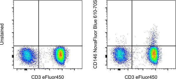
Applications Tested: This P1H12 antibody has been pre-titrated and tested by flow cytometric analysis of normal human peripheral blood cells and Human Umbilical Vein Cells (HUVEC). This can be used at 5 µL (0.02 µg) per test. A test is defined as the amount (µg) of antibody that will stain a cell sample in a final volume of 100 µL. Cell number should be determined empirically but can range from 10^5 to 10^8 cells/test.
NovaFluor dyes are not compatible with DNA intercalating viability dyes. Do not use viability dyes such as propidium iodide, 7-actinomycin D (7-AAD) and DAPI. Invitrogen LIVE/DEAD Fixable Dead Cell stains are recommended for use with NovaFluor dyes.
This NovaFluor conjugate has been updated to ship with CellBlox Plus Blocking Buffer (Cat. No. (C001T06F01)). This buffer contains formulation improvements over CellBlox. CellBlox Plus Blocking Buffer is required for optimal staining with NovaFluor conjugates and should be used in all experiments where NovaFluor conjugates are used. Whenever possible, we recommend adding CellBlox Plus Blocking Buffer to antibody cocktails/master mixes prior to combining with cells. Add 5 µL per sample (regardless of the number of NovaFluors in your panel) to use the antibody cocktail as intended. For single-color controls, use 5 µL of CellBlox Blocking Buffer per 100 µL of cell sample containing 10^3 to 10^8 cells.
NovaFluor conjugates are based on Phiton™ technology utilizing novel nucleic acid dye structures that allow for engineered fluorescent signatures with consideration for spillover and spread impacts. Learn more
Excitation: 509 nm; Emission: 614 nm; Laser: 488 nm (Blue) Laser
For Research Use Only. Not for use in diagnostic procedures. Not for resale without express authorization.
CD146 Monoclonal Antibody (P1H12), NovaFluor Blue 610-70S, eBioscience
PRODUCT DETAILS
Host: Mouse
Isotype: IgG1, kappa
Clonality: Monoclonal
Clone: P1H12
Format: NovaFluor Blue 610-70S
Reactivity: Ca, Hu, Ms, Rb
Application: Flow Cytometry
Tested Dilution: 5 µL (0.2 µg)/test
Concentration: 0.2 μg/Test
Storage: 4°C, store in dark, DO NOT FREEZE!
Formulation: PBS with 0.09% sodium azide; pH 7.2
Purification: Affinity chromatography
Data Sheet: TDS
Specific Information
Description: The monoclonal antibody P1H12 recognizes CD146 also known as MUC18, s-endo, Endo-CAM and Mel-CAM, which is a member of the Ig superfamily of proteins. The expression of CD146 is found on endothelial cells, bone marrow fibroblasts and some tumors (especially melanoma). Recently mesenchymal stromal cells and endometrial stromal cells have also been shown to express CD146. The presence of CD146 on circulating blood cells have been confined to a subset of T cells rather than circulating endothelial cells, as expression of other endothelial markers (CD31 and CD51/61) is negative. Expression can be found on activated lymphocytes. The protein is heavily glycosylated with more than 50% of the mass from carbohydrates. The antibody P1H12 has been reported to crossreact to mouse, rabbit, canine, but not rat.
Each product contains 1 vial of NovaFluor conjugate and 1 vial of CellBlox Plus Blocking Buffer .
Applications Reported: This P1H12 antibody has been reported for use in flow cytometric analysis.
Applications Tested: This P1H12 antibody has been pre-titrated and tested by flow cytometric analysis of normal human peripheral blood cells and Human Umbilical Vein Cells (HUVEC). This can be used at 5 µL (0.02 µg) per test. A test is defined as the amount (µg) of antibody that will stain a cell sample in a final volume of 100 µL. Cell number should be determined empirically but can range from 10^5 to 10^8 cells/test.
NovaFluor dyes are not compatible with DNA intercalating viability dyes. Do not use viability dyes such as propidium iodide, 7-actinomycin D (7-AAD) and DAPI. Invitrogen LIVE/DEAD Fixable Dead Cell stains are recommended for use with NovaFluor dyes.
This NovaFluor conjugate has been updated to ship with CellBlox Plus Blocking Buffer (Cat. No. (C001T06F01)). This buffer contains formulation improvements over CellBlox. CellBlox Plus Blocking Buffer is required for optimal staining with NovaFluor conjugates and should be used in all experiments where NovaFluor conjugates are used. Whenever possible, we recommend adding CellBlox Plus Blocking Buffer to antibody cocktails/master mixes prior to combining with cells. Add 5 µL per sample (regardless of the number of NovaFluors in your panel) to use the antibody cocktail as intended. For single-color controls, use 5 µL of CellBlox Blocking Buffer per 100 µL of cell sample containing 10^3 to 10^8 cells.
NovaFluor conjugates are based on Phiton™ technology utilizing novel nucleic acid dye structures that allow for engineered fluorescent signatures with consideration for spillover and spread impacts. Learn more
Excitation: 509 nm; Emission: 614 nm; Laser: 488 nm (Blue) Laser
For Research Use Only. Not for use in diagnostic procedures. Not for resale without express authorization.
Product Information
Product Information
Shipping & Returns
Shipping & Returns
Description
PRODUCT DETAILS
Host: Mouse
Isotype: IgG1, kappa
Clonality: Monoclonal
Clone: P1H12
Format: NovaFluor Blue 610-70S
Reactivity: Ca, Hu, Ms, Rb
Application: Flow Cytometry
Tested Dilution: 5 µL (0.2 µg)/test
Concentration: 0.2 μg/Test
Storage: 4°C, store in dark, DO NOT FREEZE!
Formulation: PBS with 0.09% sodium azide; pH 7.2
Purification: Affinity chromatography
Data Sheet: TDS
Specific Information
Description: The monoclonal antibody P1H12 recognizes CD146 also known as MUC18, s-endo, Endo-CAM and Mel-CAM, which is a member of the Ig superfamily of proteins. The expression of CD146 is found on endothelial cells, bone marrow fibroblasts and some tumors (especially melanoma). Recently mesenchymal stromal cells and endometrial stromal cells have also been shown to express CD146. The presence of CD146 on circulating blood cells have been confined to a subset of T cells rather than circulating endothelial cells, as expression of other endothelial markers (CD31 and CD51/61) is negative. Expression can be found on activated lymphocytes. The protein is heavily glycosylated with more than 50% of the mass from carbohydrates. The antibody P1H12 has been reported to crossreact to mouse, rabbit, canine, but not rat.
Each product contains 1 vial of NovaFluor conjugate and 1 vial of CellBlox Plus Blocking Buffer .
Applications Reported: This P1H12 antibody has been reported for use in flow cytometric analysis.
Applications Tested: This P1H12 antibody has been pre-titrated and tested by flow cytometric analysis of normal human peripheral blood cells and Human Umbilical Vein Cells (HUVEC). This can be used at 5 µL (0.02 µg) per test. A test is defined as the amount (µg) of antibody that will stain a cell sample in a final volume of 100 µL. Cell number should be determined empirically but can range from 10^5 to 10^8 cells/test.
NovaFluor dyes are not compatible with DNA intercalating viability dyes. Do not use viability dyes such as propidium iodide, 7-actinomycin D (7-AAD) and DAPI. Invitrogen LIVE/DEAD Fixable Dead Cell stains are recommended for use with NovaFluor dyes.
This NovaFluor conjugate has been updated to ship with CellBlox Plus Blocking Buffer (Cat. No. (C001T06F01)). This buffer contains formulation improvements over CellBlox. CellBlox Plus Blocking Buffer is required for optimal staining with NovaFluor conjugates and should be used in all experiments where NovaFluor conjugates are used. Whenever possible, we recommend adding CellBlox Plus Blocking Buffer to antibody cocktails/master mixes prior to combining with cells. Add 5 µL per sample (regardless of the number of NovaFluors in your panel) to use the antibody cocktail as intended. For single-color controls, use 5 µL of CellBlox Blocking Buffer per 100 µL of cell sample containing 10^3 to 10^8 cells.
NovaFluor conjugates are based on Phiton™ technology utilizing novel nucleic acid dye structures that allow for engineered fluorescent signatures with consideration for spillover and spread impacts. Learn more
Excitation: 509 nm; Emission: 614 nm; Laser: 488 nm (Blue) Laser
For Research Use Only. Not for use in diagnostic procedures. Not for resale without express authorization.


