CD140b (PDGFRB) Monoclonal Antibody (APB5), Super Bright™ 780, eBioscience™
PRODUCT DETAILS
Host: Rat
Isotype: IgG2a, kappa
Clonality: Monoclonal
Clone: APB5
Format: Super Bright™ 780
Reactivity: Mouse
Application: Flow Cytometry
Tested Dilution: 1.0 µg/test
Concentration: 0.2 mg/mL
Storage: 4° C, store in dark, DO NOT FREEZE!
Formulation: PBS, pH 7.2, containing 0.09% sodium azide
Purification: Affinity chromatography
Data Sheet: TDS
Specific Information
Description: The APB5 monoclonal antibody reacts with the mouse CD140b molecule, the beta chain of the platelet derived growth factor receptor (PDGF receptor). PDGFRb is a receptor tyrosine kinase that forms dimers on the surface upon ligand binding and phosphorylates substrates. Dimers of PDGFR consist of either homodimers of alpha/alpha, beta/beta, or heterodimers of alpha/beta and serve as a substrate for its kinase activity. CD140b is expressed by embryonic tissues and mesenchymal-derived cells of the adult mouse tissues. The PDGFR beta chain is reported to play a significant role in formation of fibrous atherosclerotic lesions.
Applications Reported: The APB5 antibody has been reported for use in flow cytometric analysis.
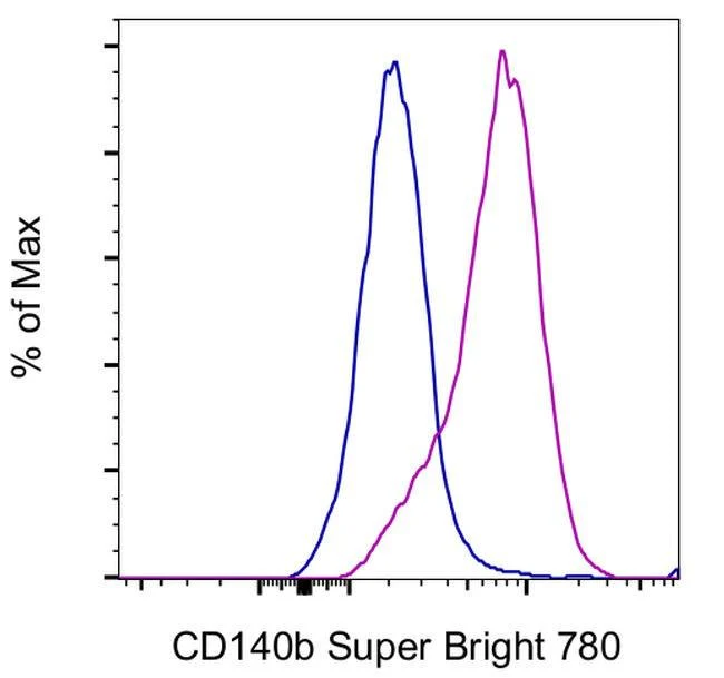
Applications Tested: The APB5 antibody has been tested by flow cytometric analysis of NIH/3T3 cells. This can be used at less than or equal to 1.0 µg per test. A test is defined as the amount (µg) of antibody that will stain a cell sample in a final volume of 100 µL. Cell number should be determined empirically but can range from 10^5 to 10^8 cells/test. It is recommended that the antibody be carefully titrated for optimal performance in the assay of interest.
Super Bright 780 is a tandem dye that can be excited with the violet laser line (405 nm) and emits at 780 nm. We recommend using a 780/60 bandpass filter. Please make sure that your instrument is capable of detecting this fluorochrome.
When using two or more Super Bright dye-conjugated antibodies in a staining panel, it is recommended to use Super Bright Complete Staining Buffer (Product # SB-4401) to minimize any non-specific polymer interactions. Please refer to the datasheet for Super Bright Staining Buffer for more information.
Light sensitivity: This tandem dye is sensitive to photo-induced oxidation. Please protect this vial and stained samples from light.
Fixation: Samples can be stored in IC Fixation Buffer (Product # 00-8222) (100 µL of cell sample + 100 µL of IC Fixation Buffer) or 1-step Fix/Lyse Solution (Product # 00-5333) for up to 3 days in the dark at 4°C with minimal impact on brightness and FRET efficiency/compensation. Some generalizations regarding fluorophore performance after fixation can be made, but clone specific performance should be determined empirically.
Excitation: 405 nm; Emission: 780 nm; Laser: Violet Laser
Super Bright Polymer Dyes are sold under license from Becton, Dickinson and Company.
For Research Use Only. Not for use in diagnostic procedures. Not for resale without express authorization.
Original: $471.00
-70%$471.00
$141.30CD140b (PDGFRB) Monoclonal Antibody (APB5), Super Bright™ 780, eBioscience™
PRODUCT DETAILS
Host: Rat
Isotype: IgG2a, kappa
Clonality: Monoclonal
Clone: APB5
Format: Super Bright™ 780
Reactivity: Mouse
Application: Flow Cytometry
Tested Dilution: 1.0 µg/test
Concentration: 0.2 mg/mL
Storage: 4° C, store in dark, DO NOT FREEZE!
Formulation: PBS, pH 7.2, containing 0.09% sodium azide
Purification: Affinity chromatography
Data Sheet: TDS
Specific Information
Description: The APB5 monoclonal antibody reacts with the mouse CD140b molecule, the beta chain of the platelet derived growth factor receptor (PDGF receptor). PDGFRb is a receptor tyrosine kinase that forms dimers on the surface upon ligand binding and phosphorylates substrates. Dimers of PDGFR consist of either homodimers of alpha/alpha, beta/beta, or heterodimers of alpha/beta and serve as a substrate for its kinase activity. CD140b is expressed by embryonic tissues and mesenchymal-derived cells of the adult mouse tissues. The PDGFR beta chain is reported to play a significant role in formation of fibrous atherosclerotic lesions.
Applications Reported: The APB5 antibody has been reported for use in flow cytometric analysis.
Applications Tested: The APB5 antibody has been tested by flow cytometric analysis of NIH/3T3 cells. This can be used at less than or equal to 1.0 µg per test. A test is defined as the amount (µg) of antibody that will stain a cell sample in a final volume of 100 µL. Cell number should be determined empirically but can range from 10^5 to 10^8 cells/test. It is recommended that the antibody be carefully titrated for optimal performance in the assay of interest.
Super Bright 780 is a tandem dye that can be excited with the violet laser line (405 nm) and emits at 780 nm. We recommend using a 780/60 bandpass filter. Please make sure that your instrument is capable of detecting this fluorochrome.
When using two or more Super Bright dye-conjugated antibodies in a staining panel, it is recommended to use Super Bright Complete Staining Buffer (Product # SB-4401) to minimize any non-specific polymer interactions. Please refer to the datasheet for Super Bright Staining Buffer for more information.
Light sensitivity: This tandem dye is sensitive to photo-induced oxidation. Please protect this vial and stained samples from light.
Fixation: Samples can be stored in IC Fixation Buffer (Product # 00-8222) (100 µL of cell sample + 100 µL of IC Fixation Buffer) or 1-step Fix/Lyse Solution (Product # 00-5333) for up to 3 days in the dark at 4°C with minimal impact on brightness and FRET efficiency/compensation. Some generalizations regarding fluorophore performance after fixation can be made, but clone specific performance should be determined empirically.
Excitation: 405 nm; Emission: 780 nm; Laser: Violet Laser
Super Bright Polymer Dyes are sold under license from Becton, Dickinson and Company.
For Research Use Only. Not for use in diagnostic procedures. Not for resale without express authorization.
Product Information
Product Information
Shipping & Returns
Shipping & Returns
Description
PRODUCT DETAILS
Host: Rat
Isotype: IgG2a, kappa
Clonality: Monoclonal
Clone: APB5
Format: Super Bright™ 780
Reactivity: Mouse
Application: Flow Cytometry
Tested Dilution: 1.0 µg/test
Concentration: 0.2 mg/mL
Storage: 4° C, store in dark, DO NOT FREEZE!
Formulation: PBS, pH 7.2, containing 0.09% sodium azide
Purification: Affinity chromatography
Data Sheet: TDS
Specific Information
Description: The APB5 monoclonal antibody reacts with the mouse CD140b molecule, the beta chain of the platelet derived growth factor receptor (PDGF receptor). PDGFRb is a receptor tyrosine kinase that forms dimers on the surface upon ligand binding and phosphorylates substrates. Dimers of PDGFR consist of either homodimers of alpha/alpha, beta/beta, or heterodimers of alpha/beta and serve as a substrate for its kinase activity. CD140b is expressed by embryonic tissues and mesenchymal-derived cells of the adult mouse tissues. The PDGFR beta chain is reported to play a significant role in formation of fibrous atherosclerotic lesions.
Applications Reported: The APB5 antibody has been reported for use in flow cytometric analysis.
Applications Tested: The APB5 antibody has been tested by flow cytometric analysis of NIH/3T3 cells. This can be used at less than or equal to 1.0 µg per test. A test is defined as the amount (µg) of antibody that will stain a cell sample in a final volume of 100 µL. Cell number should be determined empirically but can range from 10^5 to 10^8 cells/test. It is recommended that the antibody be carefully titrated for optimal performance in the assay of interest.
Super Bright 780 is a tandem dye that can be excited with the violet laser line (405 nm) and emits at 780 nm. We recommend using a 780/60 bandpass filter. Please make sure that your instrument is capable of detecting this fluorochrome.
When using two or more Super Bright dye-conjugated antibodies in a staining panel, it is recommended to use Super Bright Complete Staining Buffer (Product # SB-4401) to minimize any non-specific polymer interactions. Please refer to the datasheet for Super Bright Staining Buffer for more information.
Light sensitivity: This tandem dye is sensitive to photo-induced oxidation. Please protect this vial and stained samples from light.
Fixation: Samples can be stored in IC Fixation Buffer (Product # 00-8222) (100 µL of cell sample + 100 µL of IC Fixation Buffer) or 1-step Fix/Lyse Solution (Product # 00-5333) for up to 3 days in the dark at 4°C with minimal impact on brightness and FRET efficiency/compensation. Some generalizations regarding fluorophore performance after fixation can be made, but clone specific performance should be determined empirically.
Excitation: 405 nm; Emission: 780 nm; Laser: Violet Laser
Super Bright Polymer Dyes are sold under license from Becton, Dickinson and Company.
For Research Use Only. Not for use in diagnostic procedures. Not for resale without express authorization.