CD11b/c Monoclonal Antibody (OX42), PerCP-eFluor 710, eBioscience
PRODUCT DETAILS
Host: Mouse
Isotype: IgG2a, kappa
Clonality: Monoclonal
Clone: OX42
Format: PerCP-eFluor 710
Reactivity: Rt
Application: Flow Cytometry
Tested Dilution: 0.125 µg/test
Concentration: 0.2 mg/mL
Storage: 4°C, store in dark, DO NOT FREEZE!
Formulation: PBS with 0.09% sodium azide; pH 7.2
Purification: Affinity chromatography
Data Sheet: TDS
Specific Information
Description: This OX42 monoclonal antibody reacts with rat CD11b and CD11c. CD11b, also known as integrin alpha M or Mac-1, is a component of complement receptor 3 (CR3). CD11c, also known as integrin alpha X, is a component of complement receptor 4 (CR4). CD11b and CD11c are expressed on immune cells such as macrophages, monocytes, granulocytes, and dendritic cells. OX42 has also been shown to detect microglia in the brain, as well as cells of the liver and epidermis.
The OX42 antibody has been reported to inhibit complement-mediated rosette formation by granulocytes and macrophages. Moreover, studies demonstrate that OX42 can inhibit granulocyte aggregation.
Applications Reported: This OX42 antibody has been reported for use in flow cytometric analysis.
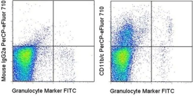
Applications Tested: This OX42 antibody has been tested by flow cytometric analysis of rat splenocytes. This can be used at less than or equal to 0.125 µg per test. A test is defined as the amount (µg) of antibody that will stain a cell sample in a final volume of 100 µL. Cell number should be determined empirically but can range from 10^5 to 10^8 cells/test. It is recommended that the antibody be carefully titrated for optimal performance in the assay of interest.
PerCP-eFluor® 710 emits at 710 nm and is excited with the blue laser (488 nm); it can be used in place of PerCP-Cyanine5.5. We recommend using a 710/50 bandpass filter, however, the 695/40 bandpass filter is an acceptable alternative. Please make sure that your instrument is capable of detecting this fluorochrome.
Fixation: Samples can be stored in IC Fixation Buffer (Product # 00-8222) (100 µL cell sample + 100 µL IC Fixation Buffer) or 1-step Fix/Lyse Solution (Product # 00-5333) for up to 3 days in the dark at 4°C with minimal impact on brightness and FRET efficiency/compensation. Some generalizations regarding fluorophore performance after fixation can be made, but clone specific performance should be determined empirically.
Excitation: 488 nm; Emission: 710 nm; Laser: Blue Laser.
Filtration: 0.2 µm post-manufacturing filtered.
For Research Use Only. Not for use in diagnostic procedures. Not for resale without express authorization.
CD11b/c Monoclonal Antibody (OX42), PerCP-eFluor 710, eBioscience
PRODUCT DETAILS
Host: Mouse
Isotype: IgG2a, kappa
Clonality: Monoclonal
Clone: OX42
Format: PerCP-eFluor 710
Reactivity: Rt
Application: Flow Cytometry
Tested Dilution: 0.125 µg/test
Concentration: 0.2 mg/mL
Storage: 4°C, store in dark, DO NOT FREEZE!
Formulation: PBS with 0.09% sodium azide; pH 7.2
Purification: Affinity chromatography
Data Sheet: TDS
Specific Information
Description: This OX42 monoclonal antibody reacts with rat CD11b and CD11c. CD11b, also known as integrin alpha M or Mac-1, is a component of complement receptor 3 (CR3). CD11c, also known as integrin alpha X, is a component of complement receptor 4 (CR4). CD11b and CD11c are expressed on immune cells such as macrophages, monocytes, granulocytes, and dendritic cells. OX42 has also been shown to detect microglia in the brain, as well as cells of the liver and epidermis.
The OX42 antibody has been reported to inhibit complement-mediated rosette formation by granulocytes and macrophages. Moreover, studies demonstrate that OX42 can inhibit granulocyte aggregation.
Applications Reported: This OX42 antibody has been reported for use in flow cytometric analysis.
Applications Tested: This OX42 antibody has been tested by flow cytometric analysis of rat splenocytes. This can be used at less than or equal to 0.125 µg per test. A test is defined as the amount (µg) of antibody that will stain a cell sample in a final volume of 100 µL. Cell number should be determined empirically but can range from 10^5 to 10^8 cells/test. It is recommended that the antibody be carefully titrated for optimal performance in the assay of interest.
PerCP-eFluor® 710 emits at 710 nm and is excited with the blue laser (488 nm); it can be used in place of PerCP-Cyanine5.5. We recommend using a 710/50 bandpass filter, however, the 695/40 bandpass filter is an acceptable alternative. Please make sure that your instrument is capable of detecting this fluorochrome.
Fixation: Samples can be stored in IC Fixation Buffer (Product # 00-8222) (100 µL cell sample + 100 µL IC Fixation Buffer) or 1-step Fix/Lyse Solution (Product # 00-5333) for up to 3 days in the dark at 4°C with minimal impact on brightness and FRET efficiency/compensation. Some generalizations regarding fluorophore performance after fixation can be made, but clone specific performance should be determined empirically.
Excitation: 488 nm; Emission: 710 nm; Laser: Blue Laser.
Filtration: 0.2 µm post-manufacturing filtered.
For Research Use Only. Not for use in diagnostic procedures. Not for resale without express authorization.
Product Information
Product Information
Shipping & Returns
Shipping & Returns
Description
PRODUCT DETAILS
Host: Mouse
Isotype: IgG2a, kappa
Clonality: Monoclonal
Clone: OX42
Format: PerCP-eFluor 710
Reactivity: Rt
Application: Flow Cytometry
Tested Dilution: 0.125 µg/test
Concentration: 0.2 mg/mL
Storage: 4°C, store in dark, DO NOT FREEZE!
Formulation: PBS with 0.09% sodium azide; pH 7.2
Purification: Affinity chromatography
Data Sheet: TDS
Specific Information
Description: This OX42 monoclonal antibody reacts with rat CD11b and CD11c. CD11b, also known as integrin alpha M or Mac-1, is a component of complement receptor 3 (CR3). CD11c, also known as integrin alpha X, is a component of complement receptor 4 (CR4). CD11b and CD11c are expressed on immune cells such as macrophages, monocytes, granulocytes, and dendritic cells. OX42 has also been shown to detect microglia in the brain, as well as cells of the liver and epidermis.
The OX42 antibody has been reported to inhibit complement-mediated rosette formation by granulocytes and macrophages. Moreover, studies demonstrate that OX42 can inhibit granulocyte aggregation.
Applications Reported: This OX42 antibody has been reported for use in flow cytometric analysis.
Applications Tested: This OX42 antibody has been tested by flow cytometric analysis of rat splenocytes. This can be used at less than or equal to 0.125 µg per test. A test is defined as the amount (µg) of antibody that will stain a cell sample in a final volume of 100 µL. Cell number should be determined empirically but can range from 10^5 to 10^8 cells/test. It is recommended that the antibody be carefully titrated for optimal performance in the assay of interest.
PerCP-eFluor® 710 emits at 710 nm and is excited with the blue laser (488 nm); it can be used in place of PerCP-Cyanine5.5. We recommend using a 710/50 bandpass filter, however, the 695/40 bandpass filter is an acceptable alternative. Please make sure that your instrument is capable of detecting this fluorochrome.
Fixation: Samples can be stored in IC Fixation Buffer (Product # 00-8222) (100 µL cell sample + 100 µL IC Fixation Buffer) or 1-step Fix/Lyse Solution (Product # 00-5333) for up to 3 days in the dark at 4°C with minimal impact on brightness and FRET efficiency/compensation. Some generalizations regarding fluorophore performance after fixation can be made, but clone specific performance should be determined empirically.
Excitation: 488 nm; Emission: 710 nm; Laser: Blue Laser.
Filtration: 0.2 µm post-manufacturing filtered.
For Research Use Only. Not for use in diagnostic procedures. Not for resale without express authorization.


