CD103 (Integrin alpha E) Monoclonal Antibody (B-Ly7), Super Bright™ 436, eBioscience™
PRODUCT DETAILS
Host: Mouse
Isotype: IgG1, kappa
Clonality: Monoclonal
Clone: B-Ly7
Format: Super Bright™ 436
Reactivity: Human
Application: Flow Cytometry
Tested Dilution: 5 µL (0.25 µg)/test
Concentration: 5 µL/Test
Storage: 4° C, store in dark, DO NOT FREEZE!
Formulation: PBS, pH 7.2, containing 0.09% sodium azide
Purification: Affinity chromatography
Data Sheet: TDS
Specific Information
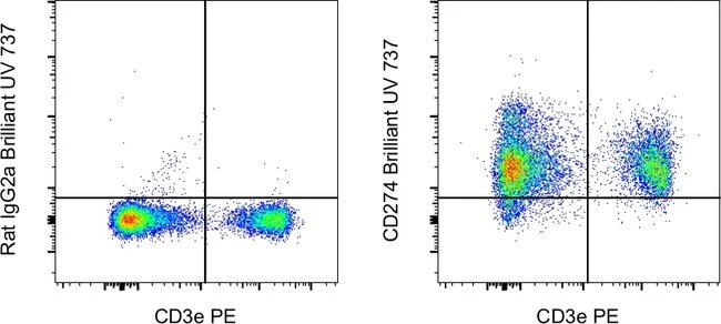
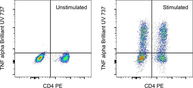
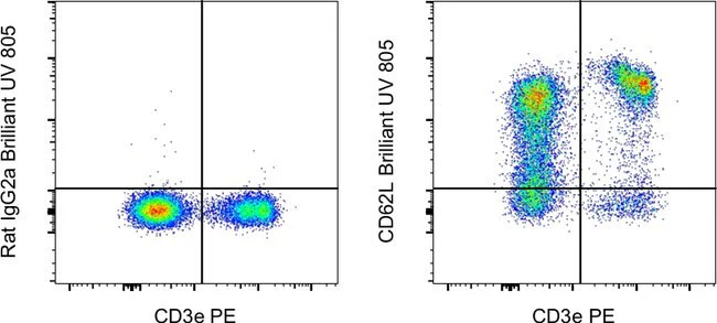
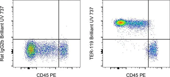
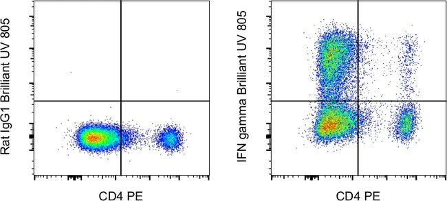
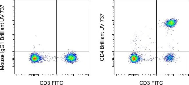
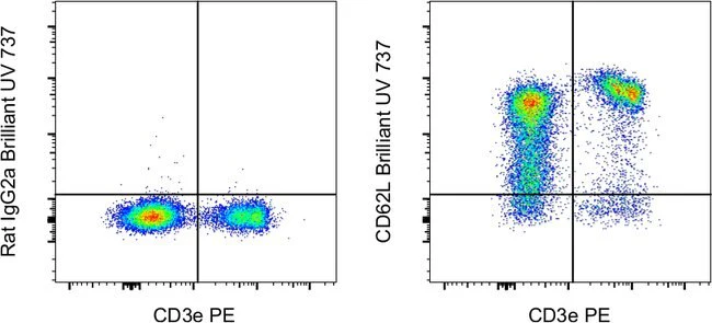
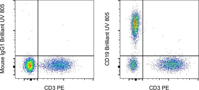
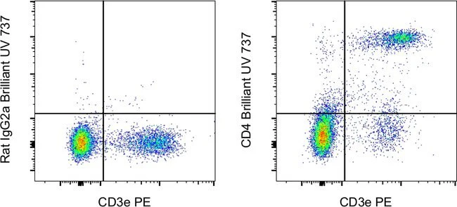
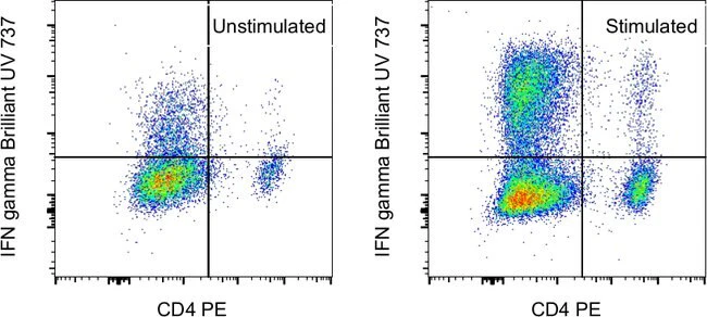
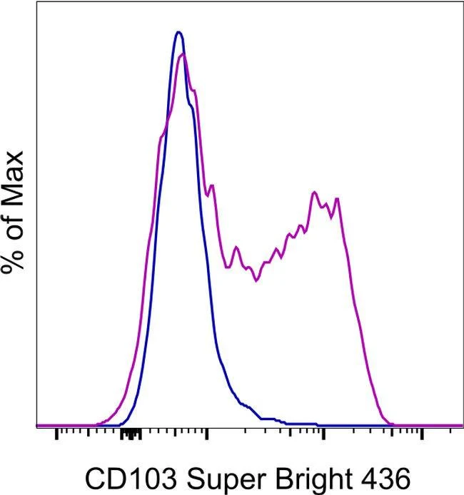
Description: The B-Ly7 monoclonal antibody reacts with human CD103, the alpha E integrin. CD103 non-covalently associates with integrin beta 7. CD103 is expressed mainly on intraepithelial lymphocytes and a small subset of peripheral lymphocytes. CD103 is also expressed by hairy cell leukemia (HCL) and by some chronic B cell lymphocytic leukemias. In vitro stimulation of human T cells with mitogens induces upregulation of CD103. Epithelial cell antigen, E-cadherin, binds to CD103 and mediates homing of lymphocytes to the intestinal epithelium.
Applications Reported: This B-Ly7 antibody has been reported for use in flow cytometric analysis.
Applications Tested: This B-Ly7 antibody has been pre-diluted and tested by flow cytometric analysis of stimulated normal human peripheral blood cells. This may be used at 5 µL (0.25 µg) per test. A test is defined as the amount (µg) of antibody that will stain a cell sample in a final volume of 100 µL. Cell number should be determined empirically but can range from 10^5 to 10^8 cells/test.
Super Bright 436 can be excited with the violet laser line (405 nm) and emits at 436 nm. We recommend using a 450/50 bandpass filter, or equivalent. Please make sure that your instrument is capable of detecting this fluorochrome.
When using two or more Super Bright dye-conjugated antibodies in a staining panel, it is recommended to use Super Bright Complete Staining Buffer (Product # SB-4401) to minimize any non-specific polymer interactions. Please refer to the datasheet for Super Bright Staining Buffer for more information.
Excitation: 405 nm; Emission: 436 nm; Laser: Violet Laser
Super Bright Polymer Dyes are sold under license from Becton, Dickinson and Company.
For Research Use Only. Not for use in diagnostic procedures. Not for resale without express authorization.
Original: $423.00
-70%$423.00
$126.90CD103 (Integrin alpha E) Monoclonal Antibody (B-Ly7), Super Bright™ 436, eBioscience™
PRODUCT DETAILS
Host: Mouse
Isotype: IgG1, kappa
Clonality: Monoclonal
Clone: B-Ly7
Format: Super Bright™ 436
Reactivity: Human
Application: Flow Cytometry
Tested Dilution: 5 µL (0.25 µg)/test
Concentration: 5 µL/Test
Storage: 4° C, store in dark, DO NOT FREEZE!
Formulation: PBS, pH 7.2, containing 0.09% sodium azide
Purification: Affinity chromatography
Data Sheet: TDS
Specific Information
Description: The B-Ly7 monoclonal antibody reacts with human CD103, the alpha E integrin. CD103 non-covalently associates with integrin beta 7. CD103 is expressed mainly on intraepithelial lymphocytes and a small subset of peripheral lymphocytes. CD103 is also expressed by hairy cell leukemia (HCL) and by some chronic B cell lymphocytic leukemias. In vitro stimulation of human T cells with mitogens induces upregulation of CD103. Epithelial cell antigen, E-cadherin, binds to CD103 and mediates homing of lymphocytes to the intestinal epithelium.
Applications Reported: This B-Ly7 antibody has been reported for use in flow cytometric analysis.
Applications Tested: This B-Ly7 antibody has been pre-diluted and tested by flow cytometric analysis of stimulated normal human peripheral blood cells. This may be used at 5 µL (0.25 µg) per test. A test is defined as the amount (µg) of antibody that will stain a cell sample in a final volume of 100 µL. Cell number should be determined empirically but can range from 10^5 to 10^8 cells/test.
Super Bright 436 can be excited with the violet laser line (405 nm) and emits at 436 nm. We recommend using a 450/50 bandpass filter, or equivalent. Please make sure that your instrument is capable of detecting this fluorochrome.
When using two or more Super Bright dye-conjugated antibodies in a staining panel, it is recommended to use Super Bright Complete Staining Buffer (Product # SB-4401) to minimize any non-specific polymer interactions. Please refer to the datasheet for Super Bright Staining Buffer for more information.
Excitation: 405 nm; Emission: 436 nm; Laser: Violet Laser
Super Bright Polymer Dyes are sold under license from Becton, Dickinson and Company.
For Research Use Only. Not for use in diagnostic procedures. Not for resale without express authorization.
Product Information
Product Information
Shipping & Returns
Shipping & Returns
Description
PRODUCT DETAILS
Host: Mouse
Isotype: IgG1, kappa
Clonality: Monoclonal
Clone: B-Ly7
Format: Super Bright™ 436
Reactivity: Human
Application: Flow Cytometry
Tested Dilution: 5 µL (0.25 µg)/test
Concentration: 5 µL/Test
Storage: 4° C, store in dark, DO NOT FREEZE!
Formulation: PBS, pH 7.2, containing 0.09% sodium azide
Purification: Affinity chromatography
Data Sheet: TDS
Specific Information
Description: The B-Ly7 monoclonal antibody reacts with human CD103, the alpha E integrin. CD103 non-covalently associates with integrin beta 7. CD103 is expressed mainly on intraepithelial lymphocytes and a small subset of peripheral lymphocytes. CD103 is also expressed by hairy cell leukemia (HCL) and by some chronic B cell lymphocytic leukemias. In vitro stimulation of human T cells with mitogens induces upregulation of CD103. Epithelial cell antigen, E-cadherin, binds to CD103 and mediates homing of lymphocytes to the intestinal epithelium.
Applications Reported: This B-Ly7 antibody has been reported for use in flow cytometric analysis.
Applications Tested: This B-Ly7 antibody has been pre-diluted and tested by flow cytometric analysis of stimulated normal human peripheral blood cells. This may be used at 5 µL (0.25 µg) per test. A test is defined as the amount (µg) of antibody that will stain a cell sample in a final volume of 100 µL. Cell number should be determined empirically but can range from 10^5 to 10^8 cells/test.
Super Bright 436 can be excited with the violet laser line (405 nm) and emits at 436 nm. We recommend using a 450/50 bandpass filter, or equivalent. Please make sure that your instrument is capable of detecting this fluorochrome.
When using two or more Super Bright dye-conjugated antibodies in a staining panel, it is recommended to use Super Bright Complete Staining Buffer (Product # SB-4401) to minimize any non-specific polymer interactions. Please refer to the datasheet for Super Bright Staining Buffer for more information.
Excitation: 405 nm; Emission: 436 nm; Laser: Violet Laser
Super Bright Polymer Dyes are sold under license from Becton, Dickinson and Company.
For Research Use Only. Not for use in diagnostic procedures. Not for resale without express authorization.