c-MAF Monoclonal Antibody (sym0F1), PE-Cyanine7, eBioscience
PRODUCT DETAILS
Host: Mouse
Isotype: IgG2b, kappa
Clonality: Monoclonal
Clone: sym0F1
Format: PE-Cyanine7
Reactivity: Hu, Ms
Application: Flow Cytometry
Tested Dilution: 0.5 µg/test
Concentration: 0.2 mg/mL
Storage: 4°C, store in dark, DO NOT FREEZE!
Formulation: PBS with 0.09% sodium azide; pH 7.2
Purification: Affinity chromatography
Data Sheet: TDS
Specific Information
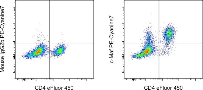
Description: The sym0F1 monoclonal antibody reacts with human and mouse c-Maf, a 42 kDa basic leucine zipper transcription factor shown to be involved in the neural, ocular and hematopoietic systems. In hematopoietic cells, it was first shown to be crucial for IL-4 expression in Th2 and was the first transcription factor believed to be Th subset-specific. More recent evidence shows that specific phospho-tyrosine residues lead to upregulation of IL-4. c-Maf has also been shown to be important to differentiation and function in both Th17 and Tfh cells. It drives expression of IL-21 in both cell types, while promoting expression of IL-23R in Th17 and CXCR5 in Tfh as well.
Applications Reported: This sym0F1 antibody has been reported for use in flow cytometric analysis.
Applications Tested: This sym0F1 antibody has been tested by flow cytometric analysis of stimulated mouse splenocytes using the Foxp3/Transcription Factor Staining Buffer Set (Product # 00-5523-00) and protocol. Please refer to "Staining Intracellular Antigens for Flow Cytometry, Protocol B: One step protocol for intracellular (nuclear) proteins" located at www.thermofisher.com/flowprotocols . This may be used at less than or equal to 0.5 µg per test. A test is defined as the amount (µg) of antibody that will stain a cell sample in a final volume of 100 µL. Cell number should be determined empirically but can range from 10^5 to 10^8 cells/test. It is recommended that the antibody be carefully titrated for optimal performance in the assay of interest.
Light sensitivity: This tandem dye is sensitive to photo-induced oxidation. Please protect this vial and stained samples from light.
Fixation: Samples can be stored in IC Fixation Buffer (Product # 00-8222-49) (100 µL of cell sample + 100 µL of IC Fixation Buffer) or 1-step Fix/Lyse Solution (Product # 00-5333-57) for up to 3 days in the dark at 4°C with minimal impact on brightness and FRET efficiency/compensation. Some generalizations regarding fluorophore performance after fixation can be made, but clone specific performance should be determined empirically.
Excitation: 488-561 nm; Emission: 775 nm; Laser: Blue Laser, Green Laser, Yellow-Green Laser.
For Research Use Only. Not for use in diagnostic procedures. Not for resale without express authorization.
Original: $495.00
-70%$495.00
$148.50c-MAF Monoclonal Antibody (sym0F1), PE-Cyanine7, eBioscience
PRODUCT DETAILS
Host: Mouse
Isotype: IgG2b, kappa
Clonality: Monoclonal
Clone: sym0F1
Format: PE-Cyanine7
Reactivity: Hu, Ms
Application: Flow Cytometry
Tested Dilution: 0.5 µg/test
Concentration: 0.2 mg/mL
Storage: 4°C, store in dark, DO NOT FREEZE!
Formulation: PBS with 0.09% sodium azide; pH 7.2
Purification: Affinity chromatography
Data Sheet: TDS
Specific Information
Description: The sym0F1 monoclonal antibody reacts with human and mouse c-Maf, a 42 kDa basic leucine zipper transcription factor shown to be involved in the neural, ocular and hematopoietic systems. In hematopoietic cells, it was first shown to be crucial for IL-4 expression in Th2 and was the first transcription factor believed to be Th subset-specific. More recent evidence shows that specific phospho-tyrosine residues lead to upregulation of IL-4. c-Maf has also been shown to be important to differentiation and function in both Th17 and Tfh cells. It drives expression of IL-21 in both cell types, while promoting expression of IL-23R in Th17 and CXCR5 in Tfh as well.
Applications Reported: This sym0F1 antibody has been reported for use in flow cytometric analysis.
Applications Tested: This sym0F1 antibody has been tested by flow cytometric analysis of stimulated mouse splenocytes using the Foxp3/Transcription Factor Staining Buffer Set (Product # 00-5523-00) and protocol. Please refer to "Staining Intracellular Antigens for Flow Cytometry, Protocol B: One step protocol for intracellular (nuclear) proteins" located at www.thermofisher.com/flowprotocols . This may be used at less than or equal to 0.5 µg per test. A test is defined as the amount (µg) of antibody that will stain a cell sample in a final volume of 100 µL. Cell number should be determined empirically but can range from 10^5 to 10^8 cells/test. It is recommended that the antibody be carefully titrated for optimal performance in the assay of interest.
Light sensitivity: This tandem dye is sensitive to photo-induced oxidation. Please protect this vial and stained samples from light.
Fixation: Samples can be stored in IC Fixation Buffer (Product # 00-8222-49) (100 µL of cell sample + 100 µL of IC Fixation Buffer) or 1-step Fix/Lyse Solution (Product # 00-5333-57) for up to 3 days in the dark at 4°C with minimal impact on brightness and FRET efficiency/compensation. Some generalizations regarding fluorophore performance after fixation can be made, but clone specific performance should be determined empirically.
Excitation: 488-561 nm; Emission: 775 nm; Laser: Blue Laser, Green Laser, Yellow-Green Laser.
For Research Use Only. Not for use in diagnostic procedures. Not for resale without express authorization.
Product Information
Product Information
Shipping & Returns
Shipping & Returns
Description
PRODUCT DETAILS
Host: Mouse
Isotype: IgG2b, kappa
Clonality: Monoclonal
Clone: sym0F1
Format: PE-Cyanine7
Reactivity: Hu, Ms
Application: Flow Cytometry
Tested Dilution: 0.5 µg/test
Concentration: 0.2 mg/mL
Storage: 4°C, store in dark, DO NOT FREEZE!
Formulation: PBS with 0.09% sodium azide; pH 7.2
Purification: Affinity chromatography
Data Sheet: TDS
Specific Information
Description: The sym0F1 monoclonal antibody reacts with human and mouse c-Maf, a 42 kDa basic leucine zipper transcription factor shown to be involved in the neural, ocular and hematopoietic systems. In hematopoietic cells, it was first shown to be crucial for IL-4 expression in Th2 and was the first transcription factor believed to be Th subset-specific. More recent evidence shows that specific phospho-tyrosine residues lead to upregulation of IL-4. c-Maf has also been shown to be important to differentiation and function in both Th17 and Tfh cells. It drives expression of IL-21 in both cell types, while promoting expression of IL-23R in Th17 and CXCR5 in Tfh as well.
Applications Reported: This sym0F1 antibody has been reported for use in flow cytometric analysis.
Applications Tested: This sym0F1 antibody has been tested by flow cytometric analysis of stimulated mouse splenocytes using the Foxp3/Transcription Factor Staining Buffer Set (Product # 00-5523-00) and protocol. Please refer to "Staining Intracellular Antigens for Flow Cytometry, Protocol B: One step protocol for intracellular (nuclear) proteins" located at www.thermofisher.com/flowprotocols . This may be used at less than or equal to 0.5 µg per test. A test is defined as the amount (µg) of antibody that will stain a cell sample in a final volume of 100 µL. Cell number should be determined empirically but can range from 10^5 to 10^8 cells/test. It is recommended that the antibody be carefully titrated for optimal performance in the assay of interest.
Light sensitivity: This tandem dye is sensitive to photo-induced oxidation. Please protect this vial and stained samples from light.
Fixation: Samples can be stored in IC Fixation Buffer (Product # 00-8222-49) (100 µL of cell sample + 100 µL of IC Fixation Buffer) or 1-step Fix/Lyse Solution (Product # 00-5333-57) for up to 3 days in the dark at 4°C with minimal impact on brightness and FRET efficiency/compensation. Some generalizations regarding fluorophore performance after fixation can be made, but clone specific performance should be determined empirically.
Excitation: 488-561 nm; Emission: 775 nm; Laser: Blue Laser, Green Laser, Yellow-Green Laser.
For Research Use Only. Not for use in diagnostic procedures. Not for resale without express authorization.


