BrdU Monoclonal Antibody (BU20A), FITC, eBioscience
PRODUCT DETAILS
Host: Mouse
Isotype: IgG1, kappa
Clonality: Monoclonal
Clone: BU20A
Format: FITC
Reactivity: Chem
Application: Flow Cytometry
Tested Dilution: 5 µL (1 µg)/test
Concentration: 5 μL/Test
Storage: 4°C, store in dark, DO NOT FREEZE!
Formulation: PBS with BSA and 0.09% sodium azide; pH 7.2
Purification: Affinity chromatography
Data Sheet: TDS
Specific Information
Description: This Bu20a monoclonal antibody reacts with 5-bromodeoxyuridine (BrdU). BrdU is a derivative of uridine that can be incorporated into DNA in place of thymidine during the S-phase of the cell cycle. Anti-BrdU can then be used to identify cells that have undergone DNA synthesis during BrdU treatment.
For staining for flow cytometric analysis, we recommend the use of the BrdU Staining Buffer Set (Product # 00-5525) and protocol.
Applications Reported: This BU20A antibody has been reported for use in intracellular staining followed by flow cytometric analysis and immunohistology staining of frozen tissue sections.
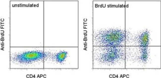
Applications Tested: This BU20A antibody has been tested by intracellular staining and flow cytometric analysis of BrdU-labeled mouse splenocytes using the Foxp3/Transcription Buffers (Product # 00-5521-00) and protocol or the BrdU Staining Buffer Set (Product # 00-5525-00) and protocol. Please see BestProtocols® Section (Staining intracellular Antigens for Flow Cytometry) for staining protocol (refer to Protocol B: One-step protocol for intracellular (nuclear) proteins). This can be used at 5µL (1 µg) per test. A test is defined as the amount (µg) of antibody that will stain a cell sample in a final volume of 100 µL. Cell number should be determined empirically but can range from 10^5 to 10^8 cells/test. It is recommended that the antibody be carefully titrated for optimal performance in the assay of interest.
BrdU labeling and staining with anti-BrdU antibody:1. Label dividing cells with 10 µM BrdU for 45 min at 37°C.2. Following the incubation, harvest the cells and wash once with 1X PBS.3. Stain surface molecules according to the Surface Staining Protocol.4. Wash in cold Flow Cytometry Staining Buffer or 1X PBS.5. Resuspend the cell pellet by pulse vortexing. Then add 1 mL of freshly prepared Foxp3 Fixation/Permeabilization Buffer (Product # 00-5523-00) to each sample. pulse vortex again.6. Incubate for 30 to 60 minutes at 2-8°C in the dark.7. Wash once with cold Flow Cytometry Staining Buffer followed by centrifugation. Decant the supernatant.8. Resuspend the cell pellet with 100 µL Flow Cytometry Staining Buffer containing 30 µg of Dnase I.9. Incubate for 1 hr at 37°C and then wash.10. Stain cells with anti-BrdU antibody for 30 min to 1 hr and then wash.10. Analyze the samples.
Excitation: 488 nm; Emission: 520 nm; Laser: Blue Laser.
Filtration: 0.2 µm post-manufacturing filtered.
For Research Use Only. Not for use in diagnostic procedures. Not for resale without express authorization.
Original: $431.00
-70%$431.00
$129.30BrdU Monoclonal Antibody (BU20A), FITC, eBioscience
PRODUCT DETAILS
Host: Mouse
Isotype: IgG1, kappa
Clonality: Monoclonal
Clone: BU20A
Format: FITC
Reactivity: Chem
Application: Flow Cytometry
Tested Dilution: 5 µL (1 µg)/test
Concentration: 5 μL/Test
Storage: 4°C, store in dark, DO NOT FREEZE!
Formulation: PBS with BSA and 0.09% sodium azide; pH 7.2
Purification: Affinity chromatography
Data Sheet: TDS
Specific Information
Description: This Bu20a monoclonal antibody reacts with 5-bromodeoxyuridine (BrdU). BrdU is a derivative of uridine that can be incorporated into DNA in place of thymidine during the S-phase of the cell cycle. Anti-BrdU can then be used to identify cells that have undergone DNA synthesis during BrdU treatment.
For staining for flow cytometric analysis, we recommend the use of the BrdU Staining Buffer Set (Product # 00-5525) and protocol.
Applications Reported: This BU20A antibody has been reported for use in intracellular staining followed by flow cytometric analysis and immunohistology staining of frozen tissue sections.
Applications Tested: This BU20A antibody has been tested by intracellular staining and flow cytometric analysis of BrdU-labeled mouse splenocytes using the Foxp3/Transcription Buffers (Product # 00-5521-00) and protocol or the BrdU Staining Buffer Set (Product # 00-5525-00) and protocol. Please see BestProtocols® Section (Staining intracellular Antigens for Flow Cytometry) for staining protocol (refer to Protocol B: One-step protocol for intracellular (nuclear) proteins). This can be used at 5µL (1 µg) per test. A test is defined as the amount (µg) of antibody that will stain a cell sample in a final volume of 100 µL. Cell number should be determined empirically but can range from 10^5 to 10^8 cells/test. It is recommended that the antibody be carefully titrated for optimal performance in the assay of interest.
BrdU labeling and staining with anti-BrdU antibody:1. Label dividing cells with 10 µM BrdU for 45 min at 37°C.2. Following the incubation, harvest the cells and wash once with 1X PBS.3. Stain surface molecules according to the Surface Staining Protocol.4. Wash in cold Flow Cytometry Staining Buffer or 1X PBS.5. Resuspend the cell pellet by pulse vortexing. Then add 1 mL of freshly prepared Foxp3 Fixation/Permeabilization Buffer (Product # 00-5523-00) to each sample. pulse vortex again.6. Incubate for 30 to 60 minutes at 2-8°C in the dark.7. Wash once with cold Flow Cytometry Staining Buffer followed by centrifugation. Decant the supernatant.8. Resuspend the cell pellet with 100 µL Flow Cytometry Staining Buffer containing 30 µg of Dnase I.9. Incubate for 1 hr at 37°C and then wash.10. Stain cells with anti-BrdU antibody for 30 min to 1 hr and then wash.10. Analyze the samples.
Excitation: 488 nm; Emission: 520 nm; Laser: Blue Laser.
Filtration: 0.2 µm post-manufacturing filtered.
For Research Use Only. Not for use in diagnostic procedures. Not for resale without express authorization.
Product Information
Product Information
Shipping & Returns
Shipping & Returns
Description
PRODUCT DETAILS
Host: Mouse
Isotype: IgG1, kappa
Clonality: Monoclonal
Clone: BU20A
Format: FITC
Reactivity: Chem
Application: Flow Cytometry
Tested Dilution: 5 µL (1 µg)/test
Concentration: 5 μL/Test
Storage: 4°C, store in dark, DO NOT FREEZE!
Formulation: PBS with BSA and 0.09% sodium azide; pH 7.2
Purification: Affinity chromatography
Data Sheet: TDS
Specific Information
Description: This Bu20a monoclonal antibody reacts with 5-bromodeoxyuridine (BrdU). BrdU is a derivative of uridine that can be incorporated into DNA in place of thymidine during the S-phase of the cell cycle. Anti-BrdU can then be used to identify cells that have undergone DNA synthesis during BrdU treatment.
For staining for flow cytometric analysis, we recommend the use of the BrdU Staining Buffer Set (Product # 00-5525) and protocol.
Applications Reported: This BU20A antibody has been reported for use in intracellular staining followed by flow cytometric analysis and immunohistology staining of frozen tissue sections.
Applications Tested: This BU20A antibody has been tested by intracellular staining and flow cytometric analysis of BrdU-labeled mouse splenocytes using the Foxp3/Transcription Buffers (Product # 00-5521-00) and protocol or the BrdU Staining Buffer Set (Product # 00-5525-00) and protocol. Please see BestProtocols® Section (Staining intracellular Antigens for Flow Cytometry) for staining protocol (refer to Protocol B: One-step protocol for intracellular (nuclear) proteins). This can be used at 5µL (1 µg) per test. A test is defined as the amount (µg) of antibody that will stain a cell sample in a final volume of 100 µL. Cell number should be determined empirically but can range from 10^5 to 10^8 cells/test. It is recommended that the antibody be carefully titrated for optimal performance in the assay of interest.
BrdU labeling and staining with anti-BrdU antibody:1. Label dividing cells with 10 µM BrdU for 45 min at 37°C.2. Following the incubation, harvest the cells and wash once with 1X PBS.3. Stain surface molecules according to the Surface Staining Protocol.4. Wash in cold Flow Cytometry Staining Buffer or 1X PBS.5. Resuspend the cell pellet by pulse vortexing. Then add 1 mL of freshly prepared Foxp3 Fixation/Permeabilization Buffer (Product # 00-5523-00) to each sample. pulse vortex again.6. Incubate for 30 to 60 minutes at 2-8°C in the dark.7. Wash once with cold Flow Cytometry Staining Buffer followed by centrifugation. Decant the supernatant.8. Resuspend the cell pellet with 100 µL Flow Cytometry Staining Buffer containing 30 µg of Dnase I.9. Incubate for 1 hr at 37°C and then wash.10. Stain cells with anti-BrdU antibody for 30 min to 1 hr and then wash.10. Analyze the samples.
Excitation: 488 nm; Emission: 520 nm; Laser: Blue Laser.
Filtration: 0.2 µm post-manufacturing filtered.
For Research Use Only. Not for use in diagnostic procedures. Not for resale without express authorization.


