beta Catenin Monoclonal Antibody (15B8), Alexa Fluor 488, eBioscience
PRODUCT DETAILS
Host: Mouse
Isotype: IgG1, kappa
Clonality: Monoclonal
Clone: 15B8
Format: Alexa Fluor 488
Reactivity: Hu, Ms
Application: Flow Cytometry
Tested Dilution: 5 µL (0.25 µg)/test
Concentration: 5 μL/Test
Storage: 4°C, store in dark, DO NOT FREEZE!
Formulation: PBS with BSA and 0.09% sodium azide; pH 7.2
Purification: Affinity chromatography
Data Sheet: TDS
Specific Information
Description: The 15B8 monoclonal antibody reacts with human and mouse beta-catenin, one member of a family of catenins, which are intracellular proteins that interact with cadherins to mediate cellular adhesion. More specifically, beta-catenin binds to the cytoplasmic tail of E-cadherin. In addition, this molecule is a component of the canonical Wnt signaling pathway. In the absence of Wnt binding its receptor, beta-catenin is phosphorylated and resides in the cytoplasm where it is eventually targeted for degradation by ubiquitination. Upon Wnt binding, beta-catenin becomes dephosphorylated, translocates to the nucleus, and modulates gene expression in partnership with the transcription factors T cell factor (TCF) and lymphocyte enhancer binding factor (LEF). Expression of beta-catenin is found in a wide variety of non-immune and immune tissues, including thymocytes and T and B lymphocytes. The Wnt and beta-catenin signaling pathway has been demonstrated to play a crucial role in the development of T, B, and hematopoietic stem cells.
Applications Reported: This 15B8 antibody has been reported for use in intracellular staining followed by flow cytometric analysis.
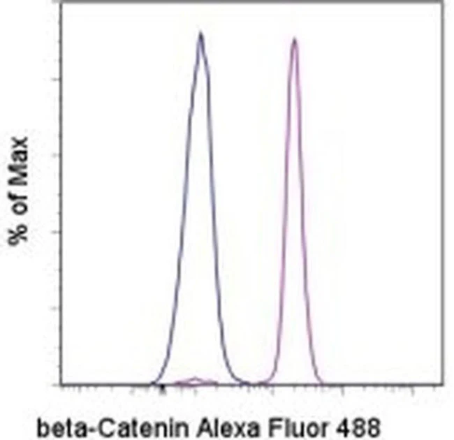
Applications Tested: This 15B8 antibody has been pre-titrated and tested by intracellular staining and flow cytometric analysis of Jurkat cell line using the Foxp3/Transcription Factor Staining Buffer Set (Product # 00-5523-00). This can be used at 5 µL (0.25 µg) per test. A test is defined as the amount (µg) of antibody that will stain a cell sample in a final volume of 100 µL. Cell number should be determined empirically but can range from 10^5 to 10^8 cells/test.
Excitation: 488 nm; Emission: 519 nm; Laser: Blue Laser.
Filtration: 0.2 µm post-manufacturing filtered.
For Research Use Only. Not for use in diagnostic procedures. Not for resale without express authorization.
beta Catenin Monoclonal Antibody (15B8), Alexa Fluor 488, eBioscience
PRODUCT DETAILS
Host: Mouse
Isotype: IgG1, kappa
Clonality: Monoclonal
Clone: 15B8
Format: Alexa Fluor 488
Reactivity: Hu, Ms
Application: Flow Cytometry
Tested Dilution: 5 µL (0.25 µg)/test
Concentration: 5 μL/Test
Storage: 4°C, store in dark, DO NOT FREEZE!
Formulation: PBS with BSA and 0.09% sodium azide; pH 7.2
Purification: Affinity chromatography
Data Sheet: TDS
Specific Information
Description: The 15B8 monoclonal antibody reacts with human and mouse beta-catenin, one member of a family of catenins, which are intracellular proteins that interact with cadherins to mediate cellular adhesion. More specifically, beta-catenin binds to the cytoplasmic tail of E-cadherin. In addition, this molecule is a component of the canonical Wnt signaling pathway. In the absence of Wnt binding its receptor, beta-catenin is phosphorylated and resides in the cytoplasm where it is eventually targeted for degradation by ubiquitination. Upon Wnt binding, beta-catenin becomes dephosphorylated, translocates to the nucleus, and modulates gene expression in partnership with the transcription factors T cell factor (TCF) and lymphocyte enhancer binding factor (LEF). Expression of beta-catenin is found in a wide variety of non-immune and immune tissues, including thymocytes and T and B lymphocytes. The Wnt and beta-catenin signaling pathway has been demonstrated to play a crucial role in the development of T, B, and hematopoietic stem cells.
Applications Reported: This 15B8 antibody has been reported for use in intracellular staining followed by flow cytometric analysis.
Applications Tested: This 15B8 antibody has been pre-titrated and tested by intracellular staining and flow cytometric analysis of Jurkat cell line using the Foxp3/Transcription Factor Staining Buffer Set (Product # 00-5523-00). This can be used at 5 µL (0.25 µg) per test. A test is defined as the amount (µg) of antibody that will stain a cell sample in a final volume of 100 µL. Cell number should be determined empirically but can range from 10^5 to 10^8 cells/test.
Excitation: 488 nm; Emission: 519 nm; Laser: Blue Laser.
Filtration: 0.2 µm post-manufacturing filtered.
For Research Use Only. Not for use in diagnostic procedures. Not for resale without express authorization.
Product Information
Product Information
Shipping & Returns
Shipping & Returns
Description
PRODUCT DETAILS
Host: Mouse
Isotype: IgG1, kappa
Clonality: Monoclonal
Clone: 15B8
Format: Alexa Fluor 488
Reactivity: Hu, Ms
Application: Flow Cytometry
Tested Dilution: 5 µL (0.25 µg)/test
Concentration: 5 μL/Test
Storage: 4°C, store in dark, DO NOT FREEZE!
Formulation: PBS with BSA and 0.09% sodium azide; pH 7.2
Purification: Affinity chromatography
Data Sheet: TDS
Specific Information
Description: The 15B8 monoclonal antibody reacts with human and mouse beta-catenin, one member of a family of catenins, which are intracellular proteins that interact with cadherins to mediate cellular adhesion. More specifically, beta-catenin binds to the cytoplasmic tail of E-cadherin. In addition, this molecule is a component of the canonical Wnt signaling pathway. In the absence of Wnt binding its receptor, beta-catenin is phosphorylated and resides in the cytoplasm where it is eventually targeted for degradation by ubiquitination. Upon Wnt binding, beta-catenin becomes dephosphorylated, translocates to the nucleus, and modulates gene expression in partnership with the transcription factors T cell factor (TCF) and lymphocyte enhancer binding factor (LEF). Expression of beta-catenin is found in a wide variety of non-immune and immune tissues, including thymocytes and T and B lymphocytes. The Wnt and beta-catenin signaling pathway has been demonstrated to play a crucial role in the development of T, B, and hematopoietic stem cells.
Applications Reported: This 15B8 antibody has been reported for use in intracellular staining followed by flow cytometric analysis.
Applications Tested: This 15B8 antibody has been pre-titrated and tested by intracellular staining and flow cytometric analysis of Jurkat cell line using the Foxp3/Transcription Factor Staining Buffer Set (Product # 00-5523-00). This can be used at 5 µL (0.25 µg) per test. A test is defined as the amount (µg) of antibody that will stain a cell sample in a final volume of 100 µL. Cell number should be determined empirically but can range from 10^5 to 10^8 cells/test.
Excitation: 488 nm; Emission: 519 nm; Laser: Blue Laser.
Filtration: 0.2 µm post-manufacturing filtered.
For Research Use Only. Not for use in diagnostic procedures. Not for resale without express authorization.


